Electrocardiogram ST Segment Elevation: Reading STEMI on EKG 2026 July
📚 Learn how to read electrocardiogram ST segment elevation on EKG, identify STEMI by lead, spot mimics, and apply ESC/AHA criteria step-by-step.

ST Elevation by the Numbers
The five numbers every reader memorizes before they ever look at a 12-lead.

Electrocardiogram ST segment elevation is the single most time-sensitive finding in cardiology. The ST segment sits between the J-point (where the QRS ends) and the start of the T-wave, and it should rest quietly on the isoelectric line — give or take half a millimetre. When that flat line lifts off the baseline by a millimetre or more in two leads that look at the same wall of the heart, you are almost certainly staring at an acute coronary occlusion, and every minute the clock keeps ticking, more cardiac muscle dies.
Most students learn STEMI as a textbook diagnosis, but in real practice it is a pattern-recognition skill. The shape of the elevation matters. The leads it appears in matter. Whether the opposite wall shows reciprocal depression matters. And the mimics — pericarditis, early repolarization, Brugada, left ventricular aneurysm, hyperkalemia, Takotsubo, Prinzmetal spasm, cocaine, pulmonary embolism, hypothermia — all matter, because activating the cath lab on a non-STEMI is expensive, and missing a real one is fatal.
This guide walks through the electrocardiogram ST-segment rules the way working EKG technicians, paramedics, and ED nurses actually use them, with the exact millimetre cut-offs, the contiguous-lead groupings, the reciprocal mirrors, and the differential checklist you should run before anyone touches a phone.
The stakes are concrete. Roughly 250,000 STEMIs occur in the United States every year, and one-year mortality remains close to 10% even with optimal reperfusion. Each 30-minute delay in opening the artery costs roughly 7.5% in absolute mortality, which is why every link in the chain — from the technician who hands the strip to the doctor, to the cath lab nurse who preps the table — exists to compress time.
Understanding what ST elevation means, what mimics it, and what to do once it shows up is therefore not a niche skill for cardiologists. It is the single most important pattern any clinician who looks at a 12-lead has to know cold.
Time Is Muscle
STEMI is a clinical emergency. The American Heart Association door-to-balloon target is 90 minutes from first medical contact to PCI. For every 30-minute delay, in-hospital mortality climbs by roughly 7.5%. If an ECG meets STEMI criteria in a patient with ischemic chest pain, the cath lab is activated before troponin results — waiting for biomarkers is a deviation from guideline care.
What Exactly Is the ST Segment?
The ST segment is the short, normally flat stretch that begins at the J-point — the junction where the QRS complex transitions into the ST — and ends where the T-wave starts to rise. Electrically, it represents the brief plateau phase of ventricular depolarization, when both ventricles are uniformly excited and no net current flows. Because there is no current, the trace should sit on the isoelectric line, which is defined by the TP segment (between the end of one T-wave and the start of the next P-wave) or the PR segment (between P and Q).
To measure ST deviation correctly, draw a baseline from the TP segment across the strip, then look at the level of the ST 0.04 to 0.08 seconds after the J-point (the so-called J+80 method recommended by the ACC/AHA). Compare that level to your baseline in millimetres — each small box on standard ECG paper is 1 mm tall at the standard 10 mm/mV calibration.
Any deviation of more than 0.5 mm above or below baseline counts as abnormal, but the diagnostic cut-offs for ischemia are stricter than that. Knowing the basics of the what is an electrocardiogram tracing matters here, because if you misidentify the isoelectric line you will misread every ST measurement that follows.
STEMI Diagnostic Thresholds (ESC/AHA 2017+)
The Formal STEMI Criteria — What Counts and What Doesn't
STEMI stands for ST Elevation Myocardial Infarction, and the diagnosis requires two things on a 12-lead. First, ST elevation that meets the millimetre thresholds above. Second, that elevation must appear in at least two anatomically contiguous leads — leads that view the same wall of the heart. A single lead showing 3 mm of STE in isolation is not a STEMI. Two adjacent leads showing 1 mm each in a patient with chest pain absolutely is.
Contiguous groupings are not arbitrary. The inferior wall is served by II, III, and aVF. The lateral wall is served by I, aVL, V5, and V6. The septum sits under V1-V2, and the anterior wall under V3-V4. Reading these electrocardiogram ecg ekg patterns is mostly about training yourself to scan in those groupings rather than lead-by-lead.
When STE appears in I and aVL but not V5-V6, it is high-lateral. When it covers V1 through V4, it is anteroseptal. The pattern tells you which coronary artery is occluded and lets the interventionalist plan the wire run before the patient is even on the table.
The ESC/AHA criteria also exclude STE caused by left bundle branch block, left ventricular hypertrophy, and electronic pacing — these conditions produce so-called secondary repolarization changes that mimic STEMI. For LBBB patients, the modified Sgarbossa criteria apply (discussed later). For LVH, the rule of thumb is that STE is considered ischemic only when it is concordant with the QRS — a discordant lift in V1-V2 is usually just strain pattern.
Localizing the Infarct by Lead Pattern
Localization is not a parlor trick — it tells the interventional team which artery to wire first and predicts the complications you should be ready for. Inferior STEMIs from RCA occlusion bring bradyarrhythmias and AV block because the RCA also feeds the SA and AV nodes in most people. Anterior STEMIs from proximal LAD occlusion bring cardiogenic shock, anterior wall akinesis on echo, and high rates of ventricular fibrillation. Lateral STEMIs from circumflex disease often have subtler ECG changes and higher rates of missed diagnosis, which is exactly why running aVL and looking for reciprocal change is so important.
Posterior infarction is the great pretender. On a standard 12-lead, it presents as horizontal ST depression in V1-V3 with prominent R-waves and upright T-waves — leads that look at the posterior wall from the front, so they show the inverse of what is really happening on the back of the heart. If you see this pattern in a patient with chest pain, place leads V7, V8, and V9 around the back at the level of V6 and the inferior tip of the scapula. STE ≥0.5 mm in any of those three confirms posterior STEMI and activates the cath lab.
How to Spot Reciprocal Depression Fast
The fastest way to look for reciprocal change is to identify the lead group with STE, then flip to its electrical opposite. Inferior STE (II, III, aVF) flips to aVL. High-lateral STE (I, aVL) flips to III. Anterior STE (V1-V4) flips to II, III, aVF.
The reciprocal lead does not always have to depress by a full millimetre — even 0.5 mm of downsloping ST depression in a single reciprocal lead, paired with millimetre-criteria STE elsewhere, is enough to confirm acute coronary occlusion in the right clinical context. Reading the rhythm and conduction context of the abnormal electrocardiogram at the same time helps — atrial flutter with variable conduction can produce pseudo-ST changes that disappear when you slow the rhythm with vagal manoeuvres.

STEMI Locations by Lead Group
Leads: II, III, aVF — STE ≥1 mm in any two of these three.
Vessel: Right coronary artery (RCA) in roughly 80% of cases; left circumflex (LCx) in the dominant-left minority.
Reciprocal: ST depression in I and aVL — a strong confirmatory finding.
Watch for: bradycardia and AV block (the RCA supplies the AV node), and right-ventricular extension. Always run V4R when you see inferior STE — RV infarction changes management (no nitrates, generous fluids).
Five Leads You Memorize Before Anything Else
The workhorse rhythm strip lead. Sees the inferior wall and shows the cleanest P-waves of any limb lead — start every interpretation here.
High-lateral view paired with lead I. Always check aVL when II/III/aVF show STE — reciprocal depression here confirms inferior STEMI.
Septal view. ST depression here with tall R-wave is the calling card of posterior infarction — do not dismiss it as normal variant.
Anterior view. The lead where Sgarbossa concordant STE ≥1 mm clinches LBBB STEMI. Also home of classic 'tombstone' elevation in massive LAD occlusion.
Right-sided lead added when inferior STE is present. STE ≥0.5 mm here means right-ventricular infarction — nitrates and morphine become dangerous.
A true STEMI almost always has a reciprocal mirror somewhere on the 12-lead. Inferior STE pairs with aVL depression. Anterior STE pairs with inferior depression. Lateral STE pairs with inferior depression. If you see ST elevation in two contiguous leads but absolutely no reciprocal change anywhere on the trace, raise your index of suspicion for a mimic — especially pericarditis, which produces diffuse STE without reciprocal depression (PR-segment depression instead).
The 10-Item ST Elevation Differential Checklist
- ✓Pericarditis — diffuse concave STE in multiple lead groups, PR-segment depression, no reciprocal change, often pleuritic chest pain worse lying down.
- ✓Early repolarization — benign normal variant in young healthy adults, notch or slur at the J-point, concave STE mostly in V2-V5, no chest pain.
- ✓Brugada syndrome — coved (type 1) STE in V1-V2 with negative T-waves, genetic sodium channel disease, risk of sudden cardiac death.
- ✓Left ventricular aneurysm — persistent STE in anterior leads weeks to months after old anterior MI, with deep Q-waves in the same leads.
- ✓Hyperkalemia — peaked T-waves first, then widened QRS, then pseudo-STEMI pattern with potassium >6.5 mmol/L.
- ✓Hypothermia — Osborn (J) waves at the QRS-ST junction when core temperature drops below 32°C, can mimic STE.
- ✓Takotsubo cardiomyopathy — stress-induced apical ballooning with anterolateral STE, often in postmenopausal women after emotional trigger, clean coronaries on angiogram.
- ✓Prinzmetal (vasospastic) angina — transient STE during coronary spasm, resolves with nitrates, classic in younger smokers and cocaine users.
- ✓Pulmonary embolism — S1Q3T3 pattern with right heart strain, occasionally STE in V1 and inferior leads from RV overload.
- ✓Left bundle branch block / paced rhythm — produces secondary repolarization STE; use modified Sgarbossa criteria to diagnose STEMI on top of LBBB.
The Ten Non-STEMI Causes of ST Elevation
Roughly one in four ECGs that meet STEMI millimetre criteria turn out to be something else after cath. The interventionalist will not thank you for a false activation, but they will absolutely never forgive a missed real STEMI, so when in doubt the rule is to err toward activation and let the diagnostic angiogram sort it out. That said, knowing the mimics tightens your specificity and helps when the patient is hemodynamically stable enough to think carefully.
Pericarditis is the classic mimic and produces a concave, saddle-shaped ST elevation that appears across multiple lead groups simultaneously (inferior and anterior at once), accompanied by PR-segment depression in the same leads and PR elevation in aVR. There are no reciprocal changes because the inflammation is global, not regional. Early repolarization shows up in athletic young adults with a fishhook-shaped J-point notch and concave STE that persists across years of ECGs.
Brugada patterns are usually picked up on screening or after a syncope work-up — the type 1 coved pattern in V1-V2 is genetic and dangerous, but it is not acute ischemia. LV aneurysm is the trap on old MI patients — persistent anterior STE with deep Q-waves represents scar tissue that never repolarizes normally, not a new occlusion.
Hyperkalemia deserves special mention because it is reversible and lethal. Potassium above 6.5 mmol/L produces tall peaked T-waves first, then PR prolongation, then QRS widening into a sine wave. Along the way it can mimic STEMI, but the broad QRS and clinical context (renal failure, missed dialysis, ACE inhibitor toxicity) point away from acute coronary occlusion. Takotsubo — stress cardiomyopathy — almost always presents in postmenopausal women after a strong emotional or physical trigger, with anterolateral STE that looks like LAD STEMI but reveals an apical ballooning pattern on ventriculogram and clean coronaries.
Prinzmetal angina produces transient STE during episodes of coronary vasospasm, resolves with sublingual nitroglycerin, and is classic in younger patients, especially smokers and people using cocaine. Cocaine-induced chest pain with STE deserves treatment as a STEMI but management differs (avoid beta-blockers, use benzodiazepines and nitrates). Pulmonary embolism produces a right-strain pattern — S1Q3T3, T-wave inversions in V1-V3, sometimes ST elevation in lead III and V1 from acute right-ventricular pressure overload.

STEMI vs NSTEMI — Two Different Diseases, Two Different Pathways
- +Complete (100%) occlusion of an epicardial coronary artery, usually from plaque rupture and thrombus.
- +ST elevation meeting millimetre criteria in 2+ contiguous leads on initial ECG.
- +Troponin will rise but cath lab activation should not wait for biomarkers.
- +Treatment pathway: emergent PCI within 90 minutes (door-to-balloon) or fibrinolysis if PCI unavailable within 120 minutes.
- +Higher in-hospital mortality (~5-7%) than NSTEMI but better long-term prognosis with timely reperfusion.
- +Anatomy on cath: single culprit lesion identifiable in >90% of cases.
- −Partial occlusion or distal embolization of an epicardial vessel — flow is reduced, not stopped.
- −ECG shows ST depression, T-wave inversion, or no acute changes — never STEMI-criteria elevation.
- −Diagnosis requires elevated troponin plus ischemic symptoms or ECG changes — biomarkers are mandatory.
- −Treatment pathway: dual antiplatelet therapy, anticoagulation, statin, beta-blocker, and angiography within 24-72 hours (risk-stratified).
- −Lower in-hospital mortality but higher one-year mortality from multivessel disease and ongoing ischemia.
- −Anatomy on cath: multivessel disease in ~60% of cases, often without a single clear culprit.
STEMI Management — The First Sixty Minutes
Once STEMI is confirmed on ECG, the protocol is well-rehearsed in every emergency department in the country. Aspirin 325 mg chewed (or 162-325 mg PR if vomiting) goes in immediately. A P2Y12 inhibitor — ticagrelor 180 mg, prasugrel 60 mg, or clopidogrel 600 mg — follows per institutional preference and bleeding risk. Anticoagulation with unfractionated heparin or bivalirudin starts pre-cath. A high-intensity statin (atorvastatin 80 mg or rosuvastatin 40 mg) is given before PCI for plaque stabilization and pleotropic benefit. Two large-bore IVs go in. Continuous cardiac monitoring is mandatory because ventricular fibrillation peaks in the first hour.
Oxygen is given only if oxygen saturation drops below 90% — routine supplemental oxygen in normoxic STEMI patients was shown by the AVOID and DETO2X-AMI trials to provide no benefit and possibly increase infarct size. Morphine is used cautiously and only for refractory pain because of the IMPACT-MI signal of increased mortality. Nitroglycerin is reasonable for ongoing pain but is contraindicated in inferior STEMI with right-ventricular involvement (where preload reduction can crash the blood pressure), in patients on phosphodiesterase inhibitors within 24-48 hours, and in profound hypotension.
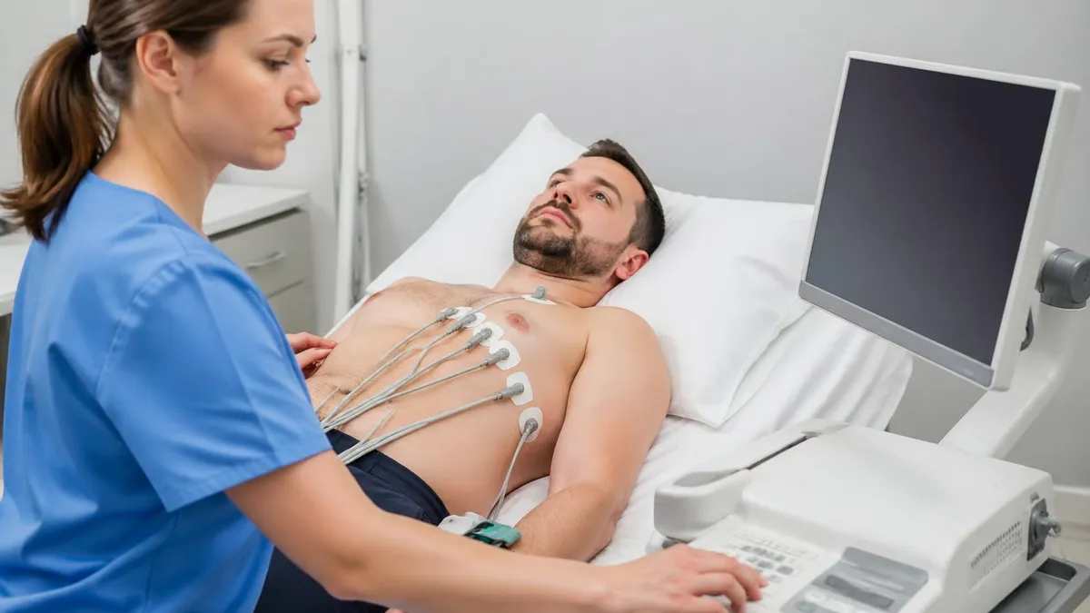
The destination is the cath lab. Door-to-balloon time — the interval from ED arrival to the moment the angioplasty balloon inflates across the culprit lesion — has a 90-minute target. Hospitals without on-site PCI must transfer to a PCI center within 120 minutes; if that is not achievable, fibrinolysis with tenecteplase or alteplase is given on-site within 30 minutes of arrival ("door-to-needle" time), then transfer for rescue or planned angiography. Many EKG technicians who pursue ekg electrocardiogram end up working in these same emergency settings, reading the strip that triggers all of this downstream activity.
Reading ST Elevation Step by Step
Working clinicians read ST elevation in the same six-step rhythm every time, and the discipline is what saves them from missed diagnoses. Step one: identify the J-point in every lead. It is the exact end of the QRS — sometimes obvious, sometimes hidden behind a slurred QRS in patients with conduction abnormality.
Step two: measure ST level 0.04 seconds (one small box) after the J-point. Some institutions use 0.08 seconds (two small boxes) — pick one and be consistent. Step three: draw the isoelectric line from the TP segment of the preceding beat and compare. ST level above the TP line = elevation; below = depression.
Step four: determine whether the elevation appears in two or more contiguous leads from the same lead group. Step five: look for reciprocal depression in the electrical mirror group — its presence dramatically raises the probability of acute coronary occlusion. Step six: correlate clinically — chest pain quality, duration, risk factors, prior ECG (if available), and initial troponin.
If the patient is asymptomatic and the elevation is concave with a J-point notch, early repolarization is far more likely than STEMI; if the patient is sweaty, gripping their chest, and has reciprocal depression, the diagnosis is made even before the lab returns. Studying focused 12 lead electrocardiogram materials reinforces these six steps until they become automatic under pressure.
The 90-Minute Door-to-Balloon Clock
Door (T+0)
First ECG (T+10)
Cath Lab Activation (T+15)
Anticoagulation + P2Y12 (T+25)
Transfer to Cath Lab (T+40)
Coronary Angiogram (T+60)
Balloon Inflation (T+90)
Left bundle branch block makes STEMI almost impossible to read on the surface 12-lead because the abnormal depolarization produces secondary STE that mimics ischemia. The Smith-modified Sgarbossa criteria — any one of: (1) concordant STE ≥1 mm in a lead with positive QRS, (2) concordant ST depression ≥1 mm in V1-V3, or (3) discordant STE ≥25% of the preceding S-wave depth in any lead — has roughly 90% specificity for acute coronary occlusion in LBBB. New or presumed-new LBBB alone is no longer considered a STEMI equivalent by current guidelines.
STEMI vs NSTEMI vs Unstable Angina — How Troponin Sorts Them
The three faces of acute coronary syndrome are separated by two simple variables: the ECG and the troponin. Unstable angina is symptomatic ischemia — chest pain at rest, new-onset crescendo angina, or post-MI angina — with no ECG changes and normal troponin. The vessel is not occluded enough to kill cells, so no troponin leaks, and the ECG looks normal or only shows non-specific T-wave flattening.
NSTEMI shares the symptoms but adds elevated troponin without ST elevation — cells are dying, the vessel is partly occluded, and the ECG typically shows ST depression or T-wave inversion. STEMI is full occlusion: chest pain plus ECG criteria for STE, with troponin rising over the following 3-6 hours but never required to make the diagnosis.
The troponin time course matters. High-sensitivity troponin assays detect myocardial necrosis as early as 1-3 hours post-onset, peak at 12-24 hours, and remain elevated for 7-14 days. A single negative high-sensitivity troponin at 3 hours, combined with a low HEART score and a normal ECG, rules out NSTEMI with >99% sensitivity and lets patients go home from the ED.
Women, the elderly, and diabetics frequently present with atypical symptoms — shortness of breath, nausea, jaw or back pain, weakness — and have lower-magnitude troponin rises, which contributes to higher rates of missed MI in these populations. An echocardiogram during or after the event confirms regional wall motion abnormalities corresponding to the infarcted territory and identifies mechanical complications like papillary muscle rupture, VSD, or free-wall rupture.
EKG Questions and Answers
Related EKG Articles

Electrocardiogram (EKG) Guide 2026: Interpretation and Certification

How Does an Electrocardiogram Work? EKG Basics Explained
EKG Certification Practice Test: Study Guide & Tips
How to Get EKG Certification: Step-by-Step Guide 2026

EKG Certification Online: How to Get Certified as an EKG Technician
EKG Certification 2026
About the Author
Educational Psychologist & Academic Test Preparation Expert
Columbia University Teachers CollegeDr. Lisa Patel holds a Doctorate in Education from Columbia University Teachers College and has spent 17 years researching standardized test design and academic assessment. She has developed preparation programs for SAT, ACT, GRE, LSAT, UCAT, and numerous professional licensing exams, helping students of all backgrounds achieve their target scores.