EEG Test Meaning: What an Electroencephalogram Measures, How It Works, and What to Expect
What is an EEG medical test? Learn how it works, what it measures, cost, side effects & duration. 🧠 Complete guide for patients.

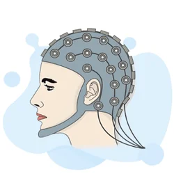
The meaning of an EEG medical test — formally known as an electroencephalogram — is a non-invasive diagnostic procedure that records the electrical activity of the brain using small metal sensors called electrodes placed on the scalp. When neurons in the brain fire, they generate tiny electrical impulses that travel in patterns. An EEG captures these patterns in real time, producing a visual record called a brain wave tracing. Neurologists and EEG technologists study these tracings to identify abnormalities linked to seizures, epilepsy, sleep disorders, brain injuries, and other neurological conditions.
An EEG test is one of the most widely ordered neurological investigations in the United States, performed in hospitals, outpatient clinics, and increasingly in ambulatory settings where patients wear a portable device for 24 to 72 hours. The test is painless and carries no radiation exposure, making it suitable for patients of all ages, from premature newborns in neonatal intensive care units to elderly patients experiencing cognitive decline or unexplained episodes of confusion.
Understanding what an EEG measures requires a basic grasp of brain electricity. The human brain contains roughly 86 billion neurons. When groups of neurons synchronize their firing, they produce oscillations at specific frequencies measured in hertz (Hz). Clinicians classify these oscillations into frequency bands: delta (0.5–4 Hz), theta (4–8 Hz), alpha (8–13 Hz), beta (13–30 Hz), and gamma (above 30 Hz). Each band correlates with distinct states of consciousness and brain function. An abnormal shift in these patterns — such as high-amplitude slow waves appearing where fast activity is expected — signals a potential problem worth investigating further.
Many patients wonder how long is an eeg test before they schedule the appointment. Standard routine EEG recordings typically last 20 to 40 minutes of actual recording time, though the full appointment including electrode placement and technologist setup can run 60 to 90 minutes. Extended recordings used when routine studies are inconclusive may last several hours. Ambulatory EEGs, worn at home, capture days of continuous data to catch infrequent or sleep-related events that a short in-clinic recording would miss entirely.
The eeg medical test is ordered for a broad range of clinical indications. The most common reason is evaluating suspected seizures or confirmed epilepsy — the EEG can show characteristic spike-and-wave discharges or focal slowing that confirms the diagnosis and helps classify the seizure type, which directly influences medication choice. Beyond seizures, EEGs are used to evaluate encephalopathy (diffuse brain dysfunction from metabolic causes, infections, or toxic exposures), assess consciousness levels in comatose patients, confirm brain death in ICU settings, and investigate sleep disorders like narcolepsy or REM sleep behavior disorder.
The clinical value of an EEG extends well beyond simply detecting epilepsy. In the emergency department, an EEG may be ordered after a first seizure, following a traumatic brain injury, or when a patient presents with unexplained altered mental status. In the neonatal ICU, continuous EEG monitoring is the gold standard for detecting subclinical seizures in critically ill newborns who cannot display the usual motor signs of a seizure. Each of these clinical contexts requires the EEG technologist to adapt their technique, electrode placement strategy, and documentation approach to capture the most diagnostically useful data.
For anyone studying to enter the EEG profession, or for patients trying to understand their upcoming procedure, grasping the fundamentals of what an EEG measures, why it is ordered, and how the results are interpreted is essential groundwork. The sections that follow break down every major aspect of the EEG test — from cost and duration to side effects and career pathways — so you arrive at your appointment or your board exam fully prepared.
EEG Test by the Numbers

Main Types of EEG Tests
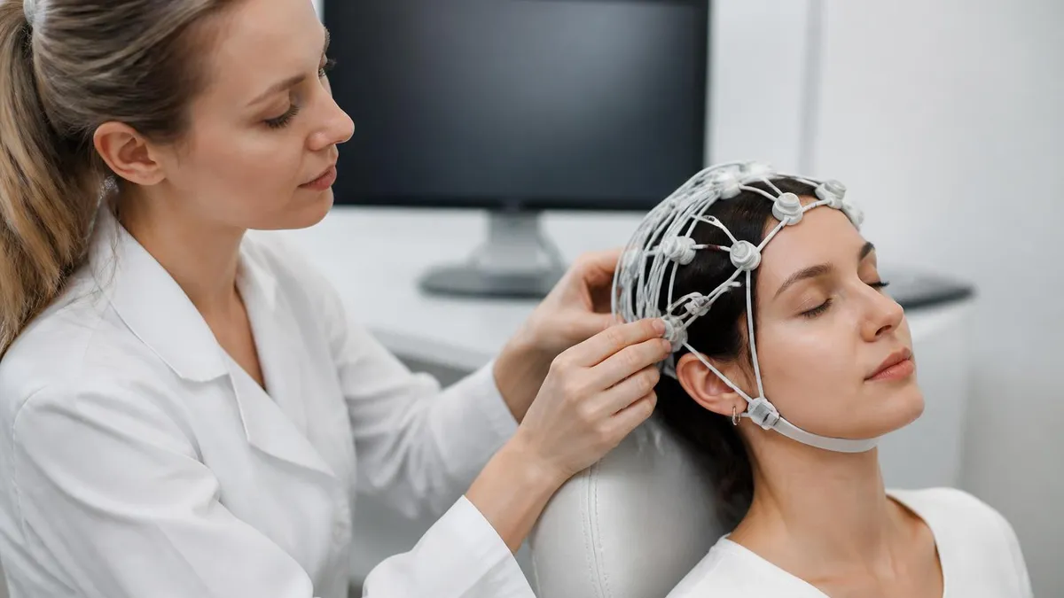
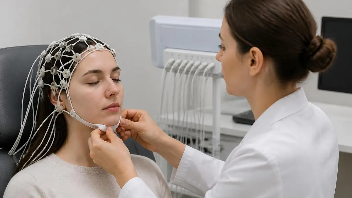
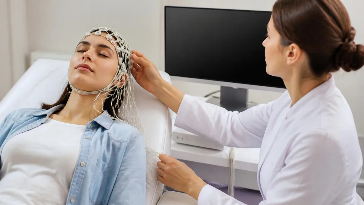
The standard 20–40 minute recording performed in a clinic or hospital. Electrodes are applied with conductive gel, the patient rests quietly, and the technologist may run activation procedures like hyperventilation or photic stimulation to provoke abnormal activity.
A portable device worn for 24 to 72 hours that continuously records brain waves during normal daily activities and sleep. Ideal for capturing infrequent events that a short routine study would miss, such as nocturnal seizures or episodes tied to specific triggers.
Combines continuous EEG recording with synchronized video monitoring in an epilepsy monitoring unit. Clinicians review EEG tracings alongside the patient's physical behavior to determine whether spells are epileptic seizures or non-epileptic events, guiding treatment decisions.
Recorded during natural sleep or after intentional sleep deprivation to capture sleep-state-specific abnormalities. Sleep activates certain epileptiform patterns and is essential for diagnosing juvenile myoclonic epilepsy and evaluating sleep architecture disorders.
Long-term monitoring in intensive care settings for critically ill patients, including those in coma or status epilepticus. Detects subclinical seizures — seizures with no visible motor signs — that can silently worsen brain injury if left untreated.
Understanding what is an eeg test at a technical level means following the signal from neuron to display. When large populations of cortical neurons fire synchronously, the summated electrical potentials are strong enough to pass through the skull, scalp tissue, and electrode gel to reach the metal sensor attached to the scalp.
Each electrode picks up the voltage difference between two points on the scalp — a principle called differential amplification — and the EEG machine amplifies these tiny signals (measured in microvolts, or millionths of a volt) by a factor of roughly one million before displaying them as wave traces on screen or paper.
The international 10-20 electrode placement system governs where each electrode sits. The system gets its name from the fact that electrode positions are spaced 10% or 20% of the total front-to-back or side-to-side skull distance apart, ensuring consistent, reproducible placement across patients and institutions. Standard positions include Fp1, Fp2 (frontal poles), F3, F4 (frontal), C3, C4 (central), P3, P4 (parietal), O1, O2 (occipital), T3, T4 (temporal), and several midline electrodes labeled with a 'z' subscript. Additional electrodes may be added to increase spatial resolution over regions of clinical interest.
Once the electrodes are in place and the recording begins, the EEG technologist asks the patient to relax and keep their eyes closed. The first segment of the recording captures the background rhythm — typically alpha waves at 8–13 Hz in a healthy, relaxed adult with eyes closed. When the patient opens their eyes, alpha activity should attenuate (decrease in amplitude), a response called alpha blocking.
Failure to produce this normal response can indicate pathology. The technologist documents these observations and any patient movements, drowsiness, or environmental artifacts in real time to help the interpreting neurologist distinguish true brain wave patterns from noise.
Activation procedures are an important part of the standard eeg test. Hyperventilation — asking the patient to breathe deeply and rapidly for three minutes — causes cerebral vasoconstriction and reduces blood flow, which can provoke absence seizure activity in susceptible patients. Photic stimulation uses a strobe light flashing at frequencies between 1 and 30 Hz to trigger photosensitive epileptiform responses.
Sleep deprivation before the recording increases the yield of the study for detecting epileptiform abnormalities, because drowsiness and early sleep stages activate interictal spikes in many epilepsy syndromes. Knowing when and how to perform these procedures is a core competency tested on the ABRET EEG Technologist credentialing examination.
EEG interpretation requires pattern recognition built through years of training. Neurologists look for several categories of abnormalities. Focal slowing — abnormally slow theta or delta waves confined to one brain region — suggests a structural lesion like a tumor, stroke, or contusion in that area. Generalized slowing involves all regions and indicates diffuse dysfunction, as seen in metabolic encephalopathies from liver or kidney failure.
Epileptiform abnormalities include interictal spikes, sharp waves, and spike-and-slow-wave complexes that appear between seizures and indicate a cortical region with abnormal excitability. Ictal patterns are the EEG changes that occur during an actual seizure, showing rhythmic evolution in frequency, amplitude, and distribution.
For patients interested in the eeg test for brain investigation pathway, it is worth knowing that a single EEG is not always conclusive. Sensitivity for detecting epileptiform abnormalities in a person with epilepsy on a single routine study is estimated at 25–56%. Repeating the study, extending the recording duration, including a sleep segment, or performing continuous monitoring substantially increases the diagnostic yield. This is why neurologists often order serial EEGs or escalate to ambulatory or inpatient video-EEG monitoring when the clinical picture strongly suggests epilepsy but the initial recording is normal.
The EEG report generated by the interpreting physician describes the background activity, any abnormalities found, whether activation procedures were performed and their results, and a clinical correlation statement linking the EEG findings to the patient's symptoms. A normal EEG does not rule out epilepsy, just as an abnormal EEG does not automatically confirm it — the EEG is one piece of a broader clinical evaluation that includes history, physical examination, and sometimes neuroimaging. Appreciating this nuance helps both patients and trainee technologists understand the EEG's role in the diagnostic process rather than viewing it as a standalone answer machine.
What Is an EEG Medical Test Used For?
Epilepsy diagnosis is the single most common reason an EEG is ordered. The test looks for interictal epileptiform discharges — spikes and sharp waves that appear between seizures — which confirm abnormal cortical excitability. Specific spike-wave patterns help classify epilepsy syndromes: 3 Hz generalized spike-wave strongly suggests childhood absence epilepsy, while hypsarrhythmia (chaotic high-amplitude activity) is the hallmark of infantile spasms. Accurate syndrome classification directly determines anticonvulsant selection and long-term prognosis.
When a patient is being evaluated for epilepsy surgery, prolonged video-EEG monitoring in an epilepsy monitoring unit localizes the seizure onset zone — the area of cortex where seizures begin. Surgeons use this information alongside MRI and other imaging to plan resection or implantation of a neurostimulation device. The EEG is indispensable in this surgical evaluation pipeline, and technologists who work in epilepsy monitoring units develop highly specialized skills in electrode placement, artifact recognition, and real-time monitoring protocols.

EEG Test Advantages and Limitations
- +Completely non-invasive — no needles, radiation, or injections required
- +Excellent temporal resolution, capturing millisecond-by-millisecond brain activity
- +Can detect seizure activity both during and between actual episodes
- +Relatively affordable compared to MRI or PET scans
- +Safe for all ages including premature newborns and elderly patients
- +Portable ambulatory versions allow real-world monitoring outside the clinic
- −Limited spatial resolution — cannot pinpoint lesion depth or precise 3D location
- −Single routine study misses epileptiform discharges in up to 50% of epilepsy cases
- −Results are highly sensitive to movement artifact, drowsiness, and patient cooperation
- −Requires skilled interpretation — misreading artifacts as pathology is a known pitfall
- −Normal EEG does not rule out epilepsy or other neurological conditions
- −Electrode application is time-consuming and gel residue can be messy for patients
How to Prepare for Your EEG Test
- ✓Wash your hair the night before or morning of the test — avoid conditioner, oils, and styling products that interfere with electrode adhesion.
- ✓Ask your doctor whether to take or skip your regular medications before the test, as some drugs affect brain wave patterns.
- ✓If a sleep-deprived EEG is ordered, stay awake the entire night before as instructed by your healthcare provider.
- ✓Avoid caffeine for at least 8 hours before the test, as it can alter brain activity and reduce diagnostic accuracy.
- ✓Eat a regular meal before your appointment — low blood sugar can cause changes in brain wave patterns.
- ✓Arrive with clean, dry hair and plan for the appointment to last 60 to 90 minutes including setup time.
- ✓Wear comfortable, loose-fitting clothing and be prepared to sit or lie still in a recliner or exam table.
- ✓Inform the EEG technologist about all medications, supplements, and any recent illnesses or sleep changes.
- ✓Tell the technologist about any fear of enclosed spaces, seizure history, or difficulty with hyperventilation exercises.
- ✓Plan for someone to drive you home if you were sleep-deprived or if your doctor anticipates you may feel tired after the test.
A Normal EEG Does Not Rule Out Epilepsy
Up to 50% of people with confirmed epilepsy have a normal routine EEG. Epileptiform discharges are often intermittent, and a 30-minute recording simply may not capture them. If your EEG is normal but your doctor still suspects seizures based on your symptoms and history, ask about repeat EEG, sleep-deprived EEG, or ambulatory monitoring — these significantly increase the diagnostic yield and should not be skipped just because the first study was unremarkable.
The eeg test cost in the United States varies substantially depending on the type of study, the setting where it is performed, and your insurance coverage. A standard routine outpatient EEG typically ranges from $200 to $900 before insurance adjustments. In a hospital outpatient department, costs may reach $1,000 to $2,000 due to facility fees that are billed separately from the professional fee charged by the interpreting neurologist.
Ambulatory EEG recordings, which last 24 to 72 hours, generally cost $500 to $1,500 for the device rental and interpretation. Prolonged inpatient video-EEG monitoring in an epilepsy monitoring unit can cost $3,000 to $10,000 or more for a multi-day admission.
Health insurance typically covers EEG testing when it is medically necessary and ordered by a licensed physician. Major commercial insurers, Medicare, and Medicaid generally reimburse EEG studies under CPT codes 95812 (EEG, 41 minutes to 1 hour), 95813 (over 1 hour), and 95816 (awake and drowsy, up to 40 minutes). Prior authorization may be required for extended or ambulatory studies. Patients without insurance or with high-deductible plans should ask the ordering facility about self-pay discounts, which often reduce the out-of-pocket cost by 30–50% compared to the billed charge.
Geographic variation in eeg test price is significant. Urban academic medical centers in high-cost-of-living states like California, New York, and Massachusetts tend to charge more than community hospitals or outpatient neurology clinics in rural areas or lower-cost states. Patients who have flexibility in where they receive care can sometimes save hundreds of dollars by choosing an independent outpatient neurology practice over a hospital-based facility, since hospital facility fees add substantially to the total bill. Calling the billing department before scheduling to ask for the cash-pay rate and any available financial assistance programs is always worthwhile.
For patients navigating financial barriers, several strategies can help reduce the cost burden. Community health centers that receive federal funding operate on a sliding-scale fee structure based on income and can perform EEGs at significantly reduced cost. University training programs affiliated with accredited EEG programs sometimes offer supervised studies at lower rates. Medical schools with neurology departments may provide access to EEG services through research protocols if the study aligns with an ongoing investigation. Advocacy organizations for epilepsy, such as the Epilepsy Foundation, maintain resources connecting patients to financial assistance programs specifically for neurological testing.
Beyond direct test costs, patients should also factor in indirect costs when budgeting for an EEG workup. If a sleep-deprived study is required, arranging child care or taking a day off work may carry its own financial impact. Transportation to the testing facility, especially for patients who cannot drive due to seizure risk, may require ride-share services or medical transport.
If the EEG leads to further testing — such as a brain MRI, which typically costs $500 to $3,000 — or a specialist consultation, the total cost of the diagnostic workup can accumulate quickly. Understanding these downstream expenses helps patients plan and advocate for themselves during insurance pre-authorization conversations.
From the perspective of the healthcare system, the EEG remains one of the most cost-effective neurological diagnostic tools available. Compared to an MRI ($500–$3,000), a CT scan ($300–$1,500), or a PET scan ($3,000–$6,000), even a hospital-based EEG is relatively affordable while providing unique information about brain function that anatomic imaging cannot supply. For patients with epilepsy who require ongoing monitoring — for example, tracking response to a new anticonvulsant or evaluating for surgical candidacy — the EEG's relatively modest cost makes serial testing feasible in a way that serial PET or MRI studies would not be.
EEG technologists who work in outpatient and hospital settings interact with billing and authorization processes regularly. Understanding CPT codes, insurance documentation requirements, and the rationale behind medical necessity criteria is part of the professional competency expected of experienced technologists. This knowledge helps them support patients navigating coverage questions and ensures the clinical documentation they generate — including detailed recording notes, artifact logs, and activation procedure records — meets the standards required for insurance reimbursement and medical record integrity.

An abnormal EEG finding does not automatically mean you have epilepsy or a serious brain condition. Many benign variants — such as wicket spikes, small sharp spikes, or 14-and-6 positive bursts — are frequently mistaken for pathology by less experienced readers. If your EEG is reported as abnormal, ask your neurologist to explain the specific finding, whether it is a definite abnormality or a normal variant, and how it correlates with your symptoms before drawing conclusions or starting treatment.
The question of eeg test side effects is one of the most common concerns patients raise before their appointment, and the reassuring answer is that a standard EEG has essentially no direct side effects. The electrodes are attached with conductive gel or paste and do not penetrate the skin. No electrical current is passed into the body — the EEG only measures, never stimulates.
After the test, patients may notice some gel residue in their hair that requires thorough shampooing to remove, and the adhesive used to hold some electrode caps may leave minor temporary redness on the scalp, but these are cosmetic inconveniences rather than medical side effects.
For a deeper exploration of eeg test side effects in specific contexts, sleep-deprived studies carry the indirect risk of drowsy driving on the way to or from the appointment. Clinicians universally advise patients who have been instructed to stay awake overnight to arrange for a driver and avoid operating any machinery for the remainder of that day. This precaution is important enough that many clinics will not perform a sleep-deprived EEG without confirming that the patient has transportation arrangements. The sleep deprivation itself — not the EEG recording — is the source of this safety concern.
Activation procedures carry slightly more nuance regarding potential effects. Hyperventilation can cause dizziness, tingling in the fingers and lips, and lightheadedness as a result of reduced carbon dioxide levels in the blood. These sensations typically resolve within 60 seconds of stopping the breathing exercise and do not represent a harmful physiological response.
Patients with certain conditions — including sickle cell disease, severe cardiovascular disease, or known moyamoya syndrome — may be advised to skip the hyperventilation phase because the transient reduction in cerebral blood flow could be harmful in their specific case. The technologist screens for these contraindications before starting the procedure.
Photic stimulation — the strobe light portion of the activation protocol — carries a very small but real risk of triggering a seizure in patients who are photosensitive. The prevalence of photosensitive epilepsy in the general population is estimated at 1 in 4,000, and the strobe procedure is specifically designed to identify this sensitivity in a controlled clinical setting where the technologist can immediately stop stimulation if a response occurs.
For the vast majority of patients, photic stimulation produces no unusual response whatsoever. The procedure uses internationally standardized stimulation frequencies to maximize diagnostic yield while minimizing the risk of a prolonged event.
Children and patients with cognitive disabilities may find the EEG setup process stressful or frightening, particularly the sensation of electrodes being applied to the scalp and the requirement to remain still. Child life specialists, parental presence, and age-appropriate distraction techniques are routinely employed to make the experience as comfortable as possible.
In rare cases where a young child cannot tolerate the procedure while awake, mild sedation may be used — typically chloral hydrate in infants or low-dose oral benzodiazepines in older children — though this is kept to a minimum because some sedatives alter the EEG background and reduce diagnostic sensitivity.
For patients with active epilepsy, the activation procedures in an EEG do carry a theoretical risk of provoking a seizure. This is actually the intended diagnostic effect when the goal is to capture ictal activity — but in a clinical recording suite, the technologist is trained to recognize seizure onset on the tracing, call for clinical assistance if a convulsion occurs, ensure patient safety by preventing falls, and document the event thoroughly.
Having a seizure during an EEG, while distressing for the patient, is not inherently dangerous in a properly equipped clinical environment and often provides the most definitive diagnostic information available.
Overall, the risk-benefit profile of an eeg medical test is exceptionally favorable. The test is so safe that it is the only neurological test routinely performed on critically ill newborns in neonatal intensive care units, premature infants whose organ systems are still developing, and patients of any age with multiple comorbidities.
For the overwhelming majority of patients referred for an outpatient routine EEG, the procedure is no more uncomfortable than having electrodes applied to the scalp, sitting quietly for 30 minutes, and then washing gel out of their hair afterward. The clinical information gained — potentially identifying a treatable seizure disorder, ruling out epilepsy, or guiding medication adjustments — vastly outweighs any inconvenience involved in the process.
For aspiring EEG technologists, understanding the full scope of practice required to perform and support eeg tests in professional settings is as important as mastering the technical recording parameters. The EEG technologist — also called an electroneurodiagnostic (END) technologist — is responsible for every step of the procedure from patient greeting and history intake through electrode application, recording, artifact management, activation procedures, and final documentation. The quality of the study rests almost entirely on the technologist's skill, patience, and ability to troubleshoot problems in real time while keeping the patient comfortable and cooperative.
Credentialing in EEG technology is managed by the American Board of Registration of Electroencephalographic and Evoked Potential Technologists (ABRET). The primary credential is the Registered EEG Technologist (R.EEG.T.) designation, which requires completing an accredited training program or equivalent on-the-job experience, passing a written examination, and maintaining continuing education credits for renewal. The examination tests competencies across electrode placement, equipment operation, normal and abnormal EEG patterns, artifact recognition, activation procedures, patient safety, and clinical documentation. Many employers in hospital neurology departments and epilepsy monitoring units require or strongly prefer R.EEG.T. credentialed candidates when hiring.
The career pathway for EEG technologists in the United States has several branching directions. Some technologists specialize in neonatal EEG monitoring, developing expertise in the unique challenges of recording from premature and term newborns whose electrode placement, waveform morphology, and normal developmental patterns differ substantially from those of adults. Others move into epilepsy monitoring unit roles, where they work closely with epileptologists on presurgical evaluations and develop advanced skills in long-term monitoring management.
Travel EEG technologists — an increasingly popular career track — work as contract professionals placed in hospitals and clinics across the country, often earning premium compensation packages that include housing stipends and travel reimbursement. For those interested in this lifestyle, exploring eeg test hospital opportunities can open doors to working in diverse clinical environments while advancing skills rapidly through varied case exposure.
Salary for EEG technologists varies by geographic location, experience level, and credential status. According to Bureau of Labor Statistics data and industry surveys, the median annual salary for electroneurodiagnostic technologists in the United States is approximately $57,000 to $64,000, with experienced credentialed technologists in high-demand markets or travel positions earning $75,000 to $90,000 or more.
ICU and epilepsy monitoring specialists who can read EEG in real time and provide immediate clinical support tend to command the highest compensation. The field is expected to grow as the aging US population increases demand for neurological diagnostic services and as continuous EEG monitoring expands beyond traditional epilepsy applications into general neurocritical care.
EEG technology programs are offered at community colleges, vocational schools, and hospital-based training programs across the United States. Program lengths typically range from 12 to 24 months for certificate programs and up to four years for associate or bachelor's degree pathways that combine EEG with broader electroneurodiagnostic technologies including evoked potentials, nerve conduction studies, and intraoperative neurophysiological monitoring.
Prospective students should verify that their chosen program is accredited by the Commission on Accreditation of Allied Health Education Programs (CAAHEP) through its Committee on Accreditation for Education in Neurodiagnostic Technology (CoA-NDT), as accreditation is required for graduates to sit for the ABRET R.EEG.T. examination.
The daily work environment of an EEG technologist is varied and clinically rich. A typical day in a busy outpatient neurology practice might include five to eight routine EEG recordings on patients ranging from young children with new-onset seizures to elderly adults being evaluated for memory problems or falls.
A hospital-based technologist on an inpatient neurology unit might split their time between bedside recordings on admitted patients, troubleshooting continuous EEG monitoring equipment in the ICU, and performing portable studies in the emergency department on patients who had a first seizure. No two days are identical, and the intellectual challenge of recognizing subtle EEG patterns and adapting technique to difficult recording conditions keeps the work engaging across a long career.
Professional development opportunities for EEG technologists have expanded significantly with the growth of online education, virtual conferences, and simulation-based training platforms. National organizations including the American Clinical Neurophysiology Society (ACNS) and the Neurodiagnostic Society publish guidelines, host annual meetings, and offer webinar series covering emerging topics in clinical EEG practice. Staying current with evolving standards — such as the ACNS guidelines on continuous EEG monitoring terminology, quantitative EEG methods, and neonatal seizure detection algorithms — is both a professional responsibility and a practical necessity in a field where the technology and clinical applications continue to advance rapidly.
EEG Questions and Answers
EEG Electroencephalography Practice Test PDF (Free Printable 2026)
Sleep EEG: How Overnight Brain Wave Testing Works and What Your Results Mean
EEG Test Cost in 2026: Prices, Insurance Coverage, and How to Save

Travel EEG Tech Jobs: The Complete Guide to Working as a Mobile Electroencephalography Technologist
EEG Tech Salary 2026: Pay by State, Experience, and Certification
About the Author
Educational Psychologist & Academic Test Preparation Expert
Columbia University Teachers CollegeDr. Lisa Patel holds a Doctorate in Education from Columbia University Teachers College and has spent 17 years researching standardized test design and academic assessment. She has developed preparation programs for SAT, ACT, GRE, LSAT, UCAT, and numerous professional licensing exams, helping students of all backgrounds achieve their target scores.