EEG Test: What It Is, What It Measures, and Why Doctors Order It 2026 July
What is an EEG test and why is it ordered? 🧠 Learn how EEG works, what conditions it diagnoses, costs, side effects, and how long it takes.

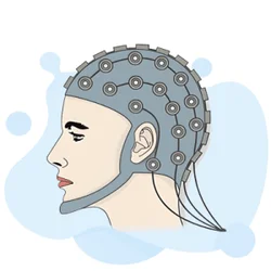
An EEG test — short for electroencephalogram — is one of the most important diagnostic tools in modern neurology. Understanding the eeg purpose starts with knowing what the test actually records: tiny electrical signals generated by millions of neurons firing in your brain. Those signals travel through your skull and scalp, where small metal electrodes pick them up and transmit them to a computer for analysis. The resulting waveform patterns tell neurologists a remarkable amount about how your brain is functioning at any given moment.
When a physician orders an EEG medical test, they are looking for abnormalities in the brain's electrical activity that cannot be seen on an MRI or CT scan. Structural imaging shows the anatomy of the brain — its shape, size, and the presence of tumors or bleeding. An EEG, by contrast, captures brain function in real time. A brain can look completely normal on an MRI and still produce dangerously abnormal electrical patterns, which is exactly why the EEG remains irreplaceable in clinical neurology more than eighty years after its invention.
Epilepsy diagnosis is far and away the most common reason patients undergo an EEG test. Seizures occur when a group of neurons fires abnormally and synchronously, creating a distinctive electrical signature called an epileptiform discharge. Even between actual seizures, many patients with epilepsy show characteristic spike-and-wave patterns or sharp waves that confirm the diagnosis. Catching these patterns allows physicians to identify the seizure type, locate where in the brain the seizures originate, and choose the most appropriate anti-seizure medication or surgical plan.
Beyond epilepsy, the EEG test is used to evaluate a wide range of neurological conditions. Physicians order it to assess altered states of consciousness, including coma and encephalopathy, where it can distinguish between reversible metabolic causes and structural brain damage. It plays a central role in confirming brain death in ICU patients. It helps diagnose sleep disorders such as narcolepsy and REM sleep behavior disorder. Researchers also rely on EEG to study cognitive processes, attention, and the neurological effects of anesthesia during surgery.
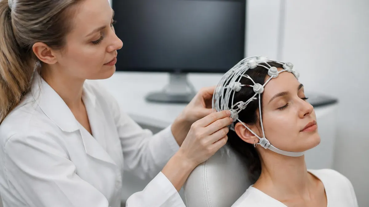
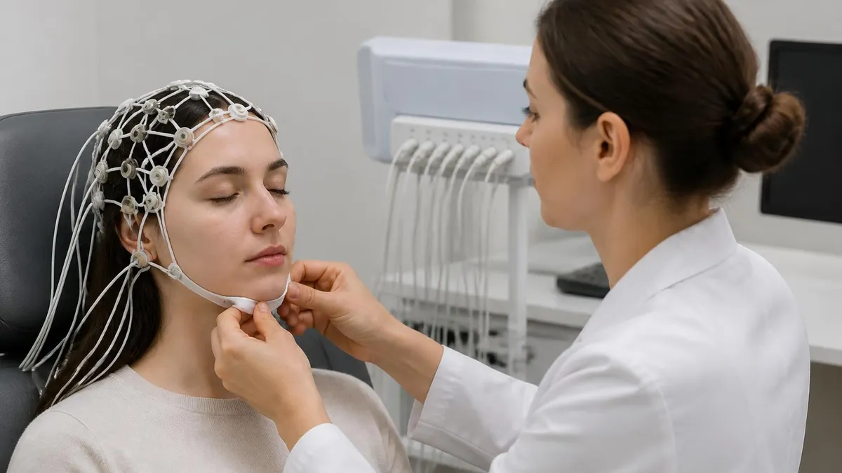
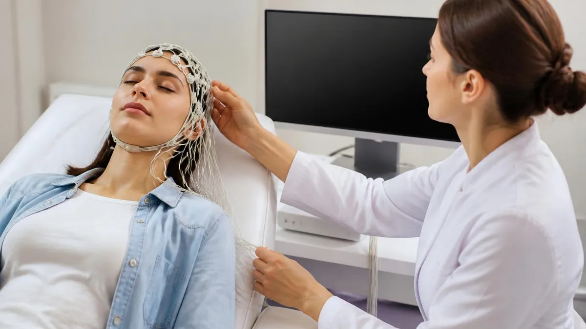
Despite being a high-technology test, the EEG is remarkably patient-friendly. No radiation is involved, no injections are required, and the electrodes record passively — they do not send any electrical current into your head. The procedure is painless, though the gel used to secure electrodes can be messy to wash out afterward. Understanding what the test involves ahead of time reduces anxiety and helps patients cooperate with the technologist, which ultimately produces a higher-quality recording for the neurologist to interpret.
EEG test cost varies significantly depending on whether you are in an inpatient or outpatient setting, which facility you use, and whether you have health insurance. A routine outpatient EEG at a hospital or freestanding neurology clinic typically runs between $200 and $700 after insurance adjustments, while the EEG test price before insurance can range from $400 to well over $2,000 at some academic medical centers. Knowing this range in advance helps patients plan financially and explore options like hospital financial assistance programs.
The field of EEG technology is constantly advancing. Digital recording systems have replaced analog paper machines, allowing technologists and neurologists to review recordings remotely and apply computerized spike-detection algorithms. Long-term video-EEG monitoring units in epilepsy centers can record continuously for days or weeks, capturing actual seizures on both video and electrical readout simultaneously. Ambulatory EEG devices let patients go home wearing a small recorder, making it possible to capture brain activity during a patient's normal daily routine — an enormous advantage for patients whose symptoms occur infrequently.
EEG Test by the Numbers

How an EEG Test Works Step by Step
Electrode Placement
Baseline Recording
Activation Procedures
Sleep or Drowsiness Phase
Signal Processing & Review
Neurologist Interpretation
The breadth of conditions that an EEG medical test can evaluate is wider than most patients realize. Epilepsy is the headline indication, but neurologists rely on EEG across the full spectrum of neurological and psychiatric disease. Understanding the specific diagnostic value the test provides in each clinical context helps patients appreciate why their physician is ordering the study and what the results will — and will not — be able to confirm.
In epilepsy care, EEG serves multiple distinct purposes at different stages of treatment. At the time of initial diagnosis, it provides objective evidence of abnormal electrical activity consistent with a seizure disorder. During treatment, serial EEGs track whether anti-seizure medications are suppressing epileptiform discharges. Before surgery, prolonged video-EEG monitoring precisely localizes the seizure onset zone so that neurosurgeons know exactly which brain region to resect. After surgery, follow-up EEGs help verify that the epileptogenic tissue has been successfully removed.
Altered consciousness is the second major domain where EEG proves indispensable. When a patient is in the intensive care unit following a stroke, cardiac arrest, traumatic brain injury, or septic encephalopathy, determining the level and nature of brain dysfunction from physical examination alone is unreliable.
Continuous EEG monitoring — where electrodes remain in place for 24 hours or longer — detects non-convulsive seizures and non-convulsive status epilepticus, conditions where dangerous electrical seizure activity is occurring without the visible shaking that most people associate with seizures. Studies at major academic medical centers estimate that between 20 and 35 percent of comatose ICU patients experience non-convulsive seizures detectable only by EEG.
Sleep medicine represents another expanding frontier for EEG. A standard polysomnography sleep study incorporates EEG channels specifically to score sleep stages: N1, N2, N3, and REM sleep each produce distinctive EEG patterns. Sleep-related epileptic disorders such as nocturnal frontal lobe epilepsy mimic other sleep conditions and can only be distinguished from them by analyzing the EEG during the events. Narcolepsy diagnosis relies on multiple sleep latency testing, which uses EEG to demonstrate pathologically rapid transitions into REM sleep.
In neonatal and pediatric neurology, EEG plays a uniquely critical role because newborns and infants may experience seizures that look nothing like the convulsions seen in adults. Subtle seizures — brief eye deviations, lip smacking, or rhythmic limb movements — are easily missed at the bedside. Continuous EEG monitoring of high-risk newborns, particularly those with hypoxic-ischemic encephalopathy, has become standard of care in Level III neonatal intensive care units across the United States because it captures seizures that would otherwise go untreated and cause permanent brain damage.
Psychiatric conditions including schizophrenia, major depression, and autism spectrum disorder all show characteristic EEG abnormalities at the population level, and quantitative EEG (qEEG) analysis is increasingly used in research settings to identify biomarkers that could guide treatment selection. While qEEG is not yet a standard clinical tool for psychiatric diagnosis, it represents one of the most promising frontiers in neuropsychiatry. The FDA has cleared one qEEG-based device as an aid in diagnosing attention deficit hyperactivity disorder in children, signaling growing regulatory acceptance of EEG-derived biomarkers.
Brain death determination is among the most solemn applications of EEG. When physicians need to confirm that all brain activity has irreversibly ceased, a properly performed EEG showing electrocerebral silence — a completely flat line with no identifiable brain electrical activity — provides important supporting evidence under the American Academy of Neurology's brain death determination guidelines. The test must be performed under strict technical standards, including high sensitivity settings and testing for electrical interference, to ensure the absence of activity is genuine and not a technical artifact obscuring residual brain function.
What Is an EEG Medical Test? Types, Duration, and Cost
There are several distinct types of EEG study, each suited to different clinical questions. A routine EEG lasts 20 to 40 minutes and is the standard first-line study for most patients. A sleep-deprived EEG asks patients to stay awake for part or all of the night before the test, making it easier to record drowsiness and light sleep states where seizure activity is more likely to emerge. Ambulatory EEG uses a small portable recorder worn for 24 to 72 hours during normal daily activities, capturing far more of the patient's typical brain activity than a brief in-clinic recording can.
Long-term video-EEG monitoring is performed in dedicated epilepsy monitoring units (EMUs) at specialized hospitals. Patients are admitted for several days to a week or longer, with continuous EEG and video recording running around the clock. Anti-seizure medications are often reduced or withdrawn to provoke seizures under controlled conditions. This approach allows neurologists to capture actual events, correlate the patient's behavior with simultaneous EEG patterns, and determine precisely where in the brain each seizure begins — information that is essential for planning epilepsy surgery.

EEG Test Advantages and Limitations
- +Non-invasive and painless — no needles, radiation, or injections required
- +Captures real-time brain function that structural imaging like MRI cannot detect
- +Extremely high temporal resolution, measuring brain activity millisecond by millisecond
- +Cost-effective first-line test compared to fMRI or PET scanning
- +Portable versions allow monitoring during normal daily activities at home
- +Can detect non-convulsive seizures invisible to bedside clinical examination
- −Limited spatial resolution — EEG cannot precisely pinpoint the 3D location of activity
- −Short routine studies may miss infrequent or sleep-dependent abnormalities
- −Electrode gel is messy and requires thorough hair washing after the test
- −Motion and muscle artifact can obscure the recording and complicate interpretation
- −Results require expert neurologist interpretation — not a point-of-care answer
- −EEG test side effects include rare skin irritation from electrode gel or paste
Preparing for Your EEG Test: 10-Step Patient Checklist
- ✓Wash your hair the night before and do NOT apply conditioner, oil, or styling products that block electrode contact.
- ✓Ask your neurologist whether to continue or temporarily stop anti-seizure medications before the test.
- ✓Follow sleep deprivation instructions exactly if ordered — staying awake until midnight or later as directed.
- ✓Eat a normal meal before the test; low blood sugar can alter background EEG rhythms.
- ✓Avoid caffeine on the morning of the test unless specifically told otherwise by your provider.
- ✓Arrive 15 to 20 minutes early to complete paperwork and allow extra time for electrode placement.
- ✓Wear comfortable, loose-fitting clothing that allows easy access to your scalp and upper body.
- ✓Bring a list of all current medications, including supplements and over-the-counter drugs, for the technologist.
- ✓Relax during the recording — muscle tension creates artifact that obscures brain signals.
- ✓Plan for someone to drive you home if you received sedation or are significantly sleep-deprived after the test.
A Normal EEG Does Not Rule Out Epilepsy
Up to 50 percent of people with confirmed epilepsy have a normal routine EEG on their first recording. Epileptiform discharges are often intermittent, appearing only during specific states such as sleep or during actual seizures. A single normal 30-minute EEG does not exclude the diagnosis — your neurologist may order a sleep-deprived study, ambulatory EEG, or long-term monitoring to increase the chance of capturing abnormalities.
Understanding how neurologists read and report EEG results empowers patients to have more informed conversations with their care team. An EEG report describes several key features of the recording, each carrying specific clinical meaning. The first is the background activity — the dominant electrical rhythm present when you are awake and relaxed.
In a healthy adult, this is the posterior dominant rhythm (PDR), a regular oscillation typically between 9 and 11 Hz that is most prominent over the back of the head and suppresses when you open your eyes. A slowed or disorganized background is one of the most common abnormal findings, indicating diffuse brain dysfunction from causes ranging from medications and metabolic disturbances to encephalitis and dementia.
Focal slowing is the term neurologists use when one region of the brain produces slower rhythms than the rest. Delta waves (below 4 Hz) or theta waves (4 to 7 Hz) appearing persistently over one hemisphere or lobe suggest a structural lesion in that area — a stroke, tumor, abscess, or area of cortical damage — because damaged neurons cannot generate the fast, organized rhythms of healthy brain tissue. When a patient has a known stroke or tumor on MRI, corresponding focal EEG slowing provides confirmation that the lesion is functionally significant, not just an incidental finding.
Epileptiform discharges are the most clinically consequential EEG abnormalities. Spikes are sharp-contoured waveforms lasting less than 70 milliseconds; sharp waves last 70 to 200 milliseconds. Both are followed by a slow wave, creating the classic spike-and-slow-wave or sharp-and-slow-wave complex. These discharge patterns are highly specific for epilepsy — they occur in roughly 2 to 4 percent of the general population but in over 90 percent of patients with well-established epilepsy. The location of discharges helps classify the epilepsy syndrome: generalized 3-Hz spike-wave is characteristic of childhood absence epilepsy, while focal temporal sharp waves suggest mesial temporal lobe epilepsy.
EEG test side effects are minimal but worth understanding before the study. The most common complaint is scalp discomfort or skin irritation at electrode sites, particularly when paste or adhesive collodion is used. Rarely, patients develop contact dermatitis from the conductive gel. Hyperventilation performed during the activation procedure regularly causes lightheadedness and tingling in the hands and lips — these sensations are normal and resolve within a minute of stopping. Photic stimulation occasionally triggers a photosensitive response in susceptible patients, which is actually a diagnostically useful finding that confirms photosensitive epilepsy rather than a complication to avoid.
Sedation, when used for pediatric EEGs or uncooperative patients, carries the standard risks associated with any sedative medication: respiratory depression, paradoxical agitation, and prolonged drowsiness. Modern EEG labs typically use chloral hydrate or dexmedetomidine under nursing supervision with pulse oximetry monitoring. General anesthesia is rarely required for EEG alone but may be used when EEG monitoring is performed during surgery. Overall, the EEG is one of the safest diagnostic procedures in medicine — its side effect profile compares favorably with even blood draws, which carry bruising and infection risks that EEG simply does not.
Quantitative EEG, or qEEG, applies mathematical analysis to the raw waveform data to extract features invisible to the naked eye. Spectral analysis calculates the power at each frequency band across all electrode sites, producing color-coded brain maps called topographic maps. Connectivity analysis measures how synchronously different brain regions communicate. Event-related potential (ERP) analysis averages hundreds of EEG epochs time-locked to a stimulus to reveal tiny brain responses hidden within background noise. These advanced techniques extend the diagnostic reach of EEG well beyond what visual inspection alone can detect.
Research applications of EEG span neuroscience, psychology, and brain-computer interface technology. Brain-computer interfaces (BCIs) use EEG signals to allow paralyzed patients to control computers, robotic arms, or speech synthesizers using only their thoughts. Neurofeedback therapy trains patients to consciously modify their own EEG patterns as a treatment for conditions including ADHD, anxiety, and PTSD.
Consumer-grade EEG headsets are now used in gaming, meditation applications, and sleep tracking, bringing EEG technology out of the clinical laboratory and into everyday life — a remarkable evolution for a test whose basic principles have not changed since Hans Berger recorded the first human EEG a century ago.

Contact your neurologist promptly if you experience a seizure within 24 hours of your EEG, especially if you had anti-seizure medications reduced before the test. Also call if you develop significant scalp redness, swelling, or a rash at electrode sites that persists more than two days, as this may indicate a contact allergy requiring treatment. Do not drive until you have fully recovered from any sedation used during the procedure.
The EEG technologist — formally titled Electroencephalographic Technologist or EEG Tech — is the clinical professional who performs the test, and understanding their role helps patients know what to expect during the procedure. EEG techs are allied health professionals trained to apply electrodes correctly, operate recording equipment, perform activation procedures safely, recognize and manage artifact, and document patient events during the recording. They work in hospital neurology departments, epilepsy monitoring units, intensive care units, outpatient neurology clinics, and research laboratories.
Becoming a certified EEG technologist requires completing an accredited EEG technology training program, which typically runs one to two years and includes both classroom instruction and clinical practicum hours. Programs cover neuroanatomy, neurophysiology, EEG instrumentation, electrode placement, pattern recognition, and patient care. Upon completing an approved program, candidates are eligible to sit for the national Registered EEG Technologist (R. EEG T.) examination administered by the American Board of Registration of Electroencephalographic and Evoked Potential Technologists (ABRET).
EEG tech salary figures for 2026 show median annual earnings around $57,000 to $75,000 depending on geographic location, level of experience, and whether the technologist holds advanced credentials such as the Registered Evoked Potential Technologist (R. EP T.) or Long-Term Monitoring Technologist (CLTM) certifications. Technologists working in major metropolitan areas, academic medical centers, or highly specialized epilepsy monitoring units tend to earn at the higher end of the range. Travel EEG tech positions — where technologists work short-term contracts at hospitals across the country — often command premium rates of $40 to $70 per hour.
The demand for EEG technologists is strong and expected to grow over the coming decade as the population ages and neurological conditions become more prevalent. Continuous EEG monitoring in ICUs has expanded dramatically over the past fifteen years as evidence mounted that non-convulsive seizures are far more common in critically ill patients than previously recognized. Each new ICU that implements a continuous EEG monitoring program needs dedicated technologists to apply electrodes, maintain recordings, and provide preliminary interpretation support around the clock, creating a steady stream of new job openings nationwide.
Advanced EEG practice includes specializations in evoked potentials — electrical responses time-locked to sensory stimuli including visual, auditory, and somatosensory tests. Visual evoked potentials (VEPs) help diagnose optic neuritis in multiple sclerosis. Brainstem auditory evoked responses (BAERs) assess hearing pathways in newborns and patients who cannot cooperate with standard audiological testing. Somatosensory evoked potentials (SSEPs) monitor spinal cord function during surgery. Technologists who master these additional modalities significantly expand their clinical utility and earning potential.
Intraoperative neurophysiological monitoring (IONM) represents a particularly specialized and high-demand application of EEG and evoked potential technology. IONM technologists accompany neurosurgeons, orthopedic surgeons, and vascular surgeons into the operating room, continuously monitoring the patient's nervous system throughout the procedure and alerting the surgical team in real time if signals change in ways that suggest imminent neurological damage. This role requires exceptional technical skill, deep physiological knowledge, and the ability to make rapid decisions under pressure — and it is compensated accordingly, with experienced IONM technologists earning $90,000 to $120,000 or more annually.
Whether you are a patient preparing for your first EEG test, a student exploring a career in neurodiagnostic technology, or a healthcare professional seeking to deepen your understanding of clinical neurophysiology, mastering EEG fundamentals opens doors across neurology, critical care, sleep medicine, pediatrics, and beyond. The resources available at PracticeTestGeeks — including practice questions, study guides, and exam simulations — provide structured, evidence-based preparation for the ABRET examinations and help aspiring technologists build the pattern recognition skills that distinguish expert EEG readers from novices.
If you are studying for the R. EEG T. examination or preparing to work in a clinical EEG lab, building strong pattern recognition skills is the single most important thing you can do. EEG interpretation is fundamentally a visual skill — neurologists and technologists learn to recognize normal and abnormal patterns the same way musicians learn to hear chords, through repeated exposure and deliberate practice. The more EEG tracings you study, the faster and more accurately you will identify artifacts, normal variants, and pathological discharges in clinical practice.
Start by thoroughly learning the normal EEG patterns for each age group and state of consciousness. The background rhythm of a healthy awake adult looks quite different from that of a healthy sleeping teenager, a normal drowsy infant, or a sedated ICU patient. Understanding what normal looks like — in all its age-dependent and state-dependent variations — is the foundation for recognizing abnormality. Many students make the mistake of jumping straight to pathological patterns without first mastering the full range of normal, which leads to over-reading artifacts and normal variants as abnormalities.
Artifact recognition is another critical competency that separates skilled EEG readers from beginners. Electroencephalograms are contaminated by dozens of non-cerebral signals during routine recording: eye blinks, eye movements, muscle tension, electrode pops, 60-Hz electrical interference, pulse artifact, sweat artifact, and movement artifact all produce waveforms that can mimic or obscure genuine brain activity. Learning to identify each artifact type by its characteristic morphology, distribution, and relationship to patient movement or environment is essential for producing accurate interpretations and for coaching patients during recording to minimize contamination.
Activation procedures deserve focused study because they are both therapeutically important and examination-testable. Hyperventilation induces cerebral vasoconstriction by lowering arterial CO2, which reduces cerebral blood flow and glucose delivery, lowering the seizure threshold in epilepsy-prone individuals. The normal hyperventilation response is generalized slowing of the background rhythm — this is a normal physiological finding, not a pathological one. Abnormal responses include focal slowing, focal epileptiform discharges, or a generalized discharge of spike-wave complexes. The distinction between the normal slowing response and a pathological response is a common examination question.
Photic stimulation activates a response called the photic driving response in normal subjects — the brain's electrical activity synchronizes to the frequency of the flashing light over the occipital regions. This is entirely normal and reflects intact visual cortex function. Pathological photosensitivity, by contrast, produces a photoparoxysmal response: generalized spike-wave or polyspike-wave discharges time-locked to the flash frequency that spread beyond the occipital region. Photoparoxysmal responses are seen in a variety of epilepsy syndromes, most commonly juvenile myoclonic epilepsy, and have significant implications for patients' safety around flickering lights.
Sleep staging is incorporated into many clinical EEG studies, and understanding the electrical hallmarks of each sleep stage is important for both technologists and neurologists. N1 sleep is characterized by the disappearance of the posterior dominant rhythm, the appearance of slow roving eye movements, and the emergence of theta activity.
N2 sleep is defined by the presence of sleep spindles (12 to 14 Hz bursts lasting 0.5 to 2 seconds) and K-complexes (high-amplitude biphasic waves). N3 or slow-wave sleep shows continuous delta activity occupying more than 20 percent of each epoch. REM sleep produces a low-amplitude mixed-frequency pattern resembling wakefulness combined with rapid eye movements and muscle atonia.
Ambulatory EEG technology has transformed the evaluation of patients whose symptoms occur infrequently or whose lifestyle makes prolonged hospital stays impractical. A modern ambulatory EEG system consists of a compact digital recorder about the size of a deck of cards, connected to a standard 10-20 electrode array and worn on a belt or shoulder harness.
Patients keep a diary of symptoms, activities, and sleep times, which is later correlated with the EEG recording to identify electrical events that coincide with reported symptoms. The combination of symptom diary and objective EEG data provides far richer diagnostic information than either source alone, making ambulatory EEG an invaluable tool in the diagnostic workup of spells, blackouts, and transient neurological events of uncertain cause.
EEG Questions and Answers
EEG Electroencephalography Practice Test PDF (Free Printable 2026)
Sleep EEG: How Overnight Brain Wave Testing Works and What Your Results Mean
EEG Test Cost in 2026: Prices, Insurance Coverage, and How to Save

Travel EEG Tech Jobs: The Complete Guide to Working as a Mobile Electroencephalography Technologist
EEG Tech Salary 2026: Pay by State, Experience, and Certification
About the Author
Educational Psychologist & Academic Test Preparation Expert
Columbia University Teachers CollegeDr. Lisa Patel holds a Doctorate in Education from Columbia University Teachers College and has spent 17 years researching standardized test design and academic assessment. She has developed preparation programs for SAT, ACT, GRE, LSAT, UCAT, and numerous professional licensing exams, helping students of all backgrounds achieve their target scores.