
CPR Flowchart: Complete Step-by-Step Guide to ACLS Algorithm, Infant CPR, and Life Support Protocols
Master the CPR flowchart, ACLS algorithm, infant CPR, and AED protocols. 🎯 Step-by-step life support guide for certification prep.

Understanding the cpr flowchart is the single most important skill any responder — trained or untrained — can carry into an emergency. The ACLS algorithm developed by the American Heart Association organizes every decision point from the moment you recognize an unresponsive victim through defibrillation, drug administration, and post-resuscitation care. Whether you are preparing for a national CPR foundation certification exam or simply want to know what to do when someone collapses in front of you, learning how each step connects to the next can mean the difference between life and death.
The foundation of every CPR flowchart begins with scene safety and responsiveness. Before touching a victim, you must confirm the environment is not dangerous to you — a rescuer who becomes a second victim helps no one. Once safety is verified, you tap the shoulders firmly and shout, check for normal breathing for no more than 10 seconds, and immediately call 911 or direct a bystander to call. These first 30 to 60 seconds of the flowchart are the most time-sensitive because brain cells begin dying within four to six minutes of cardiac arrest without adequate oxygen delivery.
The life support chain depends on compressions delivered at the correct rate and depth. For adults, the ACLS algorithm specifies a compression rate of 100 to 120 per minute and a depth of at least 2 inches but no more than 2.4 inches. Full chest recoil between compressions is mandatory — leaning on the chest prevents venous return and dramatically reduces cardiac output. Respiratory rate during CPR with an advanced airway is set at 10 breaths per minute, completely asynchronous from compressions, which allows uninterrupted chest compressions while ventilations are delivered every 6 seconds.
Infant CPR introduces significant modifications to the standard adult flowchart. For infants under one year of age, you use only two fingers centered on the sternum, just below the nipple line, and compress to a depth of approximately 1.5 inches. The compression-to-ventilation ratio for a single rescuer remains 30:2, but two trained rescuers performing infant CPR switch to a 15:2 ratio to provide more frequent ventilations for a patient population that is more likely experiencing respiratory arrest rather than primary cardiac arrest. Recognizing these differences is critical for PALS certification candidates.
What does AED stand for? An Automated External Defibrillator is a portable, computerized device that analyzes the heart's rhythm and delivers a controlled electrical shock to restore normal sinus rhythm when it detects ventricular fibrillation or pulseless ventricular tachycardia — the two shockable rhythms addressed in the ACLS algorithm.
AEDs are designed to be used by lay responders with minimal training because the device itself guides the user through each step with voice prompts and visual indicators. Every minute that passes without defibrillation in a shockable rhythm reduces survival by approximately 10 percent, making early AED use a cornerstone of the life support flowchart.
After a shock is delivered or if the rhythm is non-shockable, the flowchart directs rescuers to immediately resume high-quality compressions for two minutes before pausing to re-analyze the rhythm. This two-minute cycle continues while the team prepares IV or IO access, administers epinephrine every three to five minutes, and considers reversible causes using the Hs and Ts mnemonic — hypovolemia, hypoxia, hydrogen ion acidosis, hypo/hyperkalemia, hypothermia, tension pneumothorax, tamponade, toxins, thrombosis pulmonary, and thrombosis coronary. Identifying and correcting these causes is what separates a systematic ACLS approach from unfocused resuscitation efforts.
The recovery position is often the forgotten step at the end of the CPR flowchart. Once spontaneous circulation returns and the patient begins breathing adequately, you should place them in the lateral recovery position — on their side with the upper knee bent forward for stability — to keep the airway open and prevent aspiration if vomiting occurs. Post-cardiac-arrest care then focuses on targeted temperature management, 12-lead ECG acquisition, and transfer to a capable cardiac center, completing the full circle of the resuscitation algorithm from collapse to hospital handoff.
CPR & Cardiac Arrest by the Numbers

CPR Flowchart: Step-by-Step Sequence
Ensure Scene Safety
Check Responsiveness & Call for Help
Begin High-Quality Compressions
Attach and Use AED
Continue ACLS Algorithm Cycles
Post-Resuscitation Care & Recovery Position
The ACLS algorithm is not a single linear pathway but a series of branching decision trees designed to handle every possible cardiac arrest rhythm. The two primary branches are shockable rhythms — ventricular fibrillation (VF) and pulseless ventricular tachycardia (pVT) — and non-shockable rhythms — pulseless electrical activity (PEA) and asystole. Knowing which branch you are on within the first rhythm check determines every subsequent intervention. The national CPR foundation and the American Heart Association both structure their advanced life support curricula around this branching framework, and it appears prominently on every ACLS certification examination.
For shockable rhythms, the ACLS algorithm proceeds as follows: deliver one shock, resume compressions immediately for two minutes, check rhythm, and repeat. After the second cycle with no return of spontaneous circulation (ROSC), establish IV or IO access if not already done and administer epinephrine 1 mg every three to five minutes.
After the third cycle, consider amiodarone 300 mg IV push (with a second dose of 150 mg if needed) or lidocaine 1–1.5 mg/kg as an alternative antiarrhythmic. Each shock should be delivered at the maximum energy setting recommended by the manufacturer — typically 200 joules for biphasic devices — to maximize the probability of successful defibrillation.
Non-shockable rhythms follow a different branch that emphasizes identifying and correcting reversible causes. PEA, by definition, shows organized electrical activity on the monitor but produces no detectable pulse. Asystole shows a flat or nearly flat line. Neither benefits from defibrillation, so shocks are never appropriate for these rhythms. Instead, the team focuses on uninterrupted high-quality CPR, epinephrine every three to five minutes, and aggressive investigation of the Hs and Ts. Hypovolemia, for example, might be corrected with aggressive fluid resuscitation; tension pneumothorax requires needle decompression; tamponade requires pericardiocentesis; certain toxins may have specific antidotes.
The respiratory rate and ventilation strategy during the ACLS algorithm depend critically on whether an advanced airway is in place. Without an advanced airway, two rescuers perform 30 compressions followed by two breaths, pausing compressions for less than 10 seconds to deliver ventilations. Once an endotracheal tube, laryngeal mask airway, or supraglottic airway is secured, the ventilation rate shifts to 10 breaths per minute — one every 6 seconds — completely asynchronous from uninterrupted compressions. This change eliminates the compression pauses caused by ventilation and significantly improves coronary and cerebral perfusion pressure throughout the resuscitation.
PALS certification expands the CPR flowchart to address pediatric patients, whose cardiac arrests are far more commonly caused by respiratory failure or shock than by primary cardiac events. The pediatric ACLS equivalent begins with identifying whether the arrest is respiratory in origin, because in those cases, ventilation alone may restore spontaneous circulation without the need for chest compressions. Fluid resuscitation, glucose management, and weight-based drug dosing are all integrated into PALS algorithms that differ meaningfully from adult protocols, requiring healthcare providers who work with children to complete separate PALS certification rather than relying solely on adult ACLS training.
Drug therapy within the ACLS algorithm is tightly regulated by the flowchart. Epinephrine is the primary vasopressor used in cardiac arrest — its alpha-adrenergic effects increase aortic diastolic pressure, which directly improves coronary perfusion pressure and the likelihood of ROSC. Vasopressin was historically listed as an alternative to the first or second dose of epinephrine, but current AHA guidelines removed it from the algorithm because clinical evidence showed no benefit over epinephrine alone. Sodium bicarbonate is used only in specific circumstances such as preexisting hyperkalemia or tricyclic antidepressant toxicity, not routinely, because its indiscriminate use can worsen intracellular acidosis.
Team dynamics are as important as technical skills during an ACLS resuscitation. The team leader assigns roles clearly, communicates closed-loop (the receiver repeats the order back and confirms execution), and tracks time between epinephrine doses and rhythm checks using a timer. This structured communication prevents errors such as double-dosing medications or prolonged compression pauses. Debriefing after every resuscitation — whether successful or not — allows teams to identify performance gaps and improve adherence to the CPR flowchart in future events, which is why high-performing cardiac arrest centers treat post-resuscitation debrief as a mandatory quality improvement step.
Infant CPR, PALS Certification, and Pediatric Life Support Protocols
Infant CPR requires two-finger compressions placed on the lower half of the sternum, just below the imaginary nipple line. A single rescuer uses the two-finger technique, compressing approximately 1.5 inches at a rate of 100 to 120 per minute, with a 30:2 compression-to-ventilation ratio. Breaths must be gentle puffs covering both the mouth and nose simultaneously to avoid overinflating the tiny lungs and causing barotrauma or gastric distension that further compromises the airway.
Two trained rescuers performing infant CPR should switch to the two-thumb encircling technique, which generates higher and more consistent coronary perfusion pressures than the two-finger method. Both thumbs are placed side by side over the lower sternum while the fingers encircle and support the infant's back. The two-rescuer ratio also changes to 15:2, allowing more frequent ventilations per minute to address the typically respiratory origin of pediatric cardiac arrest. Correct technique and ratio adherence are tested directly on PALS certification examinations.

Flowchart-Based CPR Training: Structured Learning vs. Scenario-Only Practice
- +Provides a clear, memorable decision tree that reduces hesitation during high-stress emergencies
- +Aligns with national CPR foundation and AHA certification examination content, improving pass rates
- +Makes it easy to identify which branch (shockable vs. non-shockable) applies to any given rhythm
- +Helps teams coordinate roles by giving each member a predictable sequence of tasks
- +Supports self-directed study — learners can trace the flowchart without an instructor present
- +Accelerates ACLS algorithm recall during mega-code skills stations in certification courses
- −Flowcharts can feel overly rigid in real scenarios where multiple problems occur simultaneously
- −Memorizing branches without understanding underlying physiology limits problem-solving in edge cases
- −Printed flowcharts are not available during actual cardiac arrests, so recall under pressure is still required
- −Does not replace hands-on practice — knowing the flowchart does not guarantee skill proficiency
- −Pediatric flowcharts (PALS) differ enough from adult flowcharts that separate study is mandatory
- −Updates to AHA guidelines can make previously memorized flowcharts partially outdated between recertification cycles
AED Use Checklist: What Does AED Stand For and How to Use One Correctly
- ✓Confirm the scene is safe before retrieving or applying the AED to the victim.
- ✓Power on the AED by pressing the button or lifting the lid — voice prompts will guide every subsequent step.
- ✓Remove or cut away clothing to expose bare, dry chest skin before placing pads.
- ✓Attach the upper-right pad below the right collarbone and the lower-left pad on the side of the chest below the armpit.
- ✓For children under eight years or under 55 pounds, use pediatric dose-attenuating pads if available.
- ✓Allow the AED to analyze the rhythm — stop CPR and ensure no one is touching the patient during analysis.
- ✓If a shock is advised, loudly announce 'Clear!' and visually confirm no one is in contact before pressing the shock button.
- ✓Resume high-quality chest compressions immediately after the shock — do not pause to check the pulse.
- ✓Follow the AED's voice prompts for subsequent rhythm analysis cycles every two minutes.
- ✓Do not remove AED pads once applied — leave them in place for EMS personnel when they arrive on scene.
Every 1-Minute Delay in Defibrillation Cuts Survival by 10%
For victims in ventricular fibrillation, survival rates drop by approximately 10 percent for every minute that passes without defibrillation. Bystander CPR buys time by maintaining minimal perfusion, but only an AED shock can terminate VF and restore organized rhythm. This is why the ACLS algorithm and every CPR flowchart place AED attachment as a simultaneous priority with starting compressions — not a secondary step.
Life support protocols extend well beyond the minutes of active cardiac arrest and include the entire continuum from bystander response through emergency department stabilization and intensive care unit management. The post-cardiac-arrest care bundle — sometimes called the fifth link in the chain of survival — addresses the complex physiology that follows return of spontaneous circulation. The myocardium is stunned, cerebral autoregulation is impaired, and the systemic ischemia-reperfusion response triggers inflammation that can cause multi-organ dysfunction if not proactively managed. Understanding this phase of the CPR flowchart is essential for ACLS-level providers.
Targeted temperature management (TTM), previously called therapeutic hypothermia, is one of the most evidence-based interventions in post-resuscitation care. Current AHA guidelines recommend preventing fever (temperature above 37.5°C) and actively managing temperature in comatose survivors of cardiac arrest, though the specific target temperature between 32°C and 36°C is now left to clinical judgment based on institutional protocols and individual patient factors. The rationale is that elevated temperature after cardiac arrest accelerates neurological injury through increased metabolic demand, excitotoxicity, and inflammatory signaling in already-vulnerable brain tissue.
A 12-lead electrocardiogram should be obtained as soon as possible after ROSC to identify ST-elevation myocardial infarction (STEMI), which requires emergent coronary angiography and percutaneous coronary intervention (PCI). Multiple studies have demonstrated that timely PCI in post-cardiac-arrest STEMI patients dramatically improves both survival and neurological outcomes. Even in the absence of STEMI on the 12-lead, many post-arrest patients have significant coronary artery disease that contributed to the arrest, making cardiology consultation and early cath lab evaluation important considerations in the overall life support continuum.
Hemodynamic optimization after ROSC targets a mean arterial pressure above 65 mmHg to ensure adequate cerebral and organ perfusion. Many post-arrest patients are profoundly hypotensive due to myocardial stunning, vasodilation, and the vasoplegic syndrome that can accompany the post-arrest state. Norepinephrine is typically the vasopressor of choice for maintaining blood pressure, while dobutamine may be added if cardiac output is critically reduced. Continuous arterial line monitoring and, in many institutions, pulmonary artery catheter or point-of-care echocardiography guide these decisions in real time.
Oxygenation targets after resuscitation are deliberately conservative. While oxygen supplementation is essential during active CPR, high-dose oxygen after ROSC can be harmful because hyperoxia generates free radicals that worsen reperfusion injury in the brain and heart. Current guidelines recommend titrating inspired oxygen to maintain SpO2 between 92 and 98 percent — avoiding both hypoxia and hyperoxia. Similarly, ventilation targets aim for normocapnia (PaCO2 35–45 mmHg) because both hypocapnia-induced cerebral vasoconstriction and hypercapnia-related cerebral vasodilation can worsen outcomes in the post-arrest brain.
Glucose management is another important but often overlooked component of post-cardiac-arrest care. Hyperglycemia after cardiac arrest is common due to the catecholamine surge and stress response, and it is independently associated with worse neurological outcomes. Most protocols target blood glucose between 140 and 180 mg/dL using insulin infusions, while avoiding tight glycemic control below 140 mg/dL because hypoglycemia is equally harmful to the recovering brain and difficult to detect in comatose, sedated patients. Frequent glucose checks — every one to two hours initially — are standard practice in post-arrest ICU care.
Neuroprognostication — determining which patients will recover meaningful neurological function — is one of the most ethically and clinically challenging aspects of post-cardiac-arrest care. Guidelines recommend waiting at least 72 hours after rewarming from TTM before making prognostic assessments, because brain recovery can continue for days and early neurological examinations are unreliable in the setting of sedation, hypothermia, and metabolic derangement. Multimodal prognostication using CT brain imaging, EEG, somatosensory evoked potentials, and serum biomarkers like neuron-specific enolase provides the most accurate picture and informs family counseling and goals-of-care discussions.

One of the most common and dangerous errors during cardiac arrest resuscitation is pausing chest compressions to check for a pulse before the rhythm check window. Guidelines specify that pulse checks should occur only during the planned two-minute rhythm analysis pause, and only if an organized rhythm is detected on the monitor. Interrupting compressions at any other time dramatically reduces coronary perfusion pressure and decreases the chance of successful defibrillation and ROSC.
Preparing for CPR and ACLS certification examinations requires more than reading a textbook — it demands active recall of the flowchart under simulated pressure. The most effective study method is to draw the ACLS algorithm from memory repeatedly until every branch point is automatic. Begin with the universal adult cardiac arrest algorithm, then add the specific shockable and non-shockable branches, drug dosing intervals, and post-ROSC care steps. Once the adult algorithm is solid, transition to the PALS pathways and infant CPR modifications, noting specifically where the protocols diverge from adult standards.
Practice quizzes are invaluable for identifying gaps in flowchart knowledge before the actual certification exam. Many candidates discover during quiz practice that they know the major branches but struggle with specific details — the correct joule setting for pediatric defibrillation, the exact milligram dose of amiodarone after the third shock, or the precise respiratory rate for a ventilated patient with an advanced airway in place. These details matter on certification examinations from the national CPR foundation and AHA, and they matter even more in real resuscitations where precision directly affects patient outcomes.
The mega-code station is the defining skills assessment in ACLS certification courses. You will be assigned a team leader role and walked through a simulated cardiac arrest scenario while evaluators assess your ability to direct team members, maintain the flowchart sequence, communicate closed-loop, minimize compression interruptions, interpret monitor rhythms, and make correct drug decisions in real time. Candidates who have memorized the flowchart as a passive list often struggle when asked to make dynamic decisions under time pressure, while those who have practiced active recall and scenario-based decision-making consistently outperform their peers in the mega-code environment.
One practical tip for the mega-code is to verbalize your reasoning aloud even when it feels unnecessary. Saying "Rhythm check shows VF — charging to maximum energy — everyone clear" accomplishes two things simultaneously: it demonstrates to the evaluator that you have correctly identified the rhythm and are following the shockable branch of the ACLS algorithm, and it cues your team to stop touching the patient and prepare for the shock. This habit of narrating the flowchart step you are executing also reduces errors because it forces deliberate, sequential thinking instead of reactive improvisation.
PALS certification candidates should pay particular attention to the systematic pediatric assessment framework that precedes any intervention. The pediatric assessment triangle — evaluating appearance, work of breathing, and circulation to the skin — allows you to categorize the child's condition in the first 30 seconds without touching them.
A child who is alert, crying vigorously, and pink is in a dramatically different physiological state than one who is limp, silent, and mottled, and the CPR flowchart response differs accordingly. Mastering this assessment framework is what distinguishes PALS-certified providers from those who simply know adult ACLS and attempt to apply it to pediatric patients.
Time management during ACLS and PALS courses is a commonly underestimated challenge. Most two-day ACLS courses pack written exams, skills stations, and mega-code simulations into an intensive schedule that leaves little room for review. Arriving prepared — having studied the flowcharts, practiced rhythm interpretation on free online ECG databases, and completed the pre-course self-assessment — ensures that course time is spent reinforcing and applying knowledge rather than learning it from scratch. Many candidates who struggle with certification courses do so because they underestimated the preparation required, not because the material itself is beyond their capability.
After you earn your CPR or ACLS certification, maintaining proficiency between renewal cycles requires deliberate practice. Review the flowcharts quarterly, participate in hospital-based mock codes if you work in a clinical setting, and use free online resources including the quiz-based practice tools on sites like PracticeTestGeeks to keep your recall sharp.
Certification renewal intervals — typically two years for ACLS and BLS — are set based on evidence that skill decay occurs within 6 to 12 months of initial training, making ongoing practice not just optional but clinically essential. Your patients deserve a provider who can execute the CPR flowchart flawlessly the first time, every time.
Practical mastery of the CPR flowchart in real-world settings depends on automaticity — the ability to execute each step without consciously thinking about what comes next. Automaticity is built through deliberate repetition, not through reading or passive review. Physical practice on a CPR manikin, ideally with real-time feedback devices that measure compression rate, depth, and recoil, is the gold standard for developing and maintaining psychomotor skill. Studies comparing trained rescuers show that those who practice on feedback manikins deliver higher-quality CPR than those who trained without feedback, and this advantage persists at the six-month skill-retention assessment.
Understanding the rationale behind each step in the CPR flowchart makes the sequence far easier to remember and adapt when real situations deviate from the textbook scenario. Compressions maintain forward blood flow because they directly increase intrathoracic pressure and compress the heart between the sternum and spine; full recoil is mandatory because negative intrathoracic pressure during the recoil phase drives venous return that fills the heart for the next compression.
Knowing this mechanism explains why leaning on the chest — which prevents full recoil — is so damaging, and it makes the guideline memorable rather than an arbitrary rule to be recited on an exam.
When practicing for ACLS certification, focus particular attention on the transition points in the flowchart — the moments when the algorithm branches based on new information. The transition from CPR to rhythm check, from rhythm check to shock decision or drug administration, and from active resuscitation to post-ROSC care are all points where cognitive overload and time pressure can cause errors.
Practicing these transitions explicitly — running through scenarios where you must quickly decide whether a rhythm is shockable, organize the team for a shock, and immediately resume compressions — builds the pattern recognition that transforms the flowchart from a checklist into an internalized decision framework.
Position recovery is another clinical skill that should be practiced alongside active CPR steps. The lateral recovery position requires you to kneel beside the patient, extend the arm nearest you at a right angle to the body, bring the far arm across the chest and hold the back of the hand against the patient's cheek, then use your free hand to pull the far knee up and roll the patient toward you onto their side.
The upper leg is bent forward at the knee to prevent the patient from rolling prone, and the head is tilted back gently to maintain airway patency. Practicing this positioning on a willing colleague or manikin before you need it in an emergency ensures you can execute it smoothly when a patient regains spontaneous circulation.
The CPR cell phone repair mnemonic is a popular community memory device that has nothing to do with electronics — it is a reminder that just as you would not attempt to repair a broken phone without the right tools and knowledge, you should not attempt resuscitation without having current CPR training.
While it is a lighthearted analogy, the underlying message is serious: CPR skill without current training and practice creates false confidence that can lead to errors including incorrect compression rate, inadequate depth, improper hand placement, and failure to minimize interruptions. Keeping your certification current and your skills sharp is not bureaucratic compliance — it is the foundation of effective emergency response.
For healthcare providers who encounter cardiac arrest infrequently, simulation-based training is the most evidence-supported method for maintaining ACLS competency between certification renewals. High-fidelity simulation centers allow teams to practice the full resuscitation sequence — including medication preparation, airway management, defibrillation, and post-ROSC care handoffs — in a realistic environment with debriefing from expert instructors. Research consistently shows that simulation-trained teams perform better on technical and non-technical skills during actual cardiac arrests compared to teams who rely solely on periodic recertification without interval practice.
Building a habit of reviewing the CPR flowchart before high-risk situations — shift changes in the ICU, new patient admissions in the emergency department, or the start of a surgical procedure — takes only a few minutes and significantly reduces cognitive load when an actual arrest occurs.
Some experienced providers keep a laminated flowchart card in their scrub pocket not because they cannot remember the steps but as a backup check during the rare but real moments when stress overwhelms working memory. These practical habits, combined with regular quiz-based self-assessment and hands-on skills practice, ensure that your ability to deliver life-saving care remains sharp, accurate, and ready to deploy whenever a patient needs it most.
CPR Questions and Answers
How Long Does CPR Certification Last? 2026 Renewal Guide
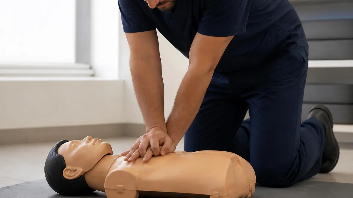
Extracorporeal CPR (ECPR): Complete Guide to Mechanical Circulatory Support During Cardiac Arrest in 2026
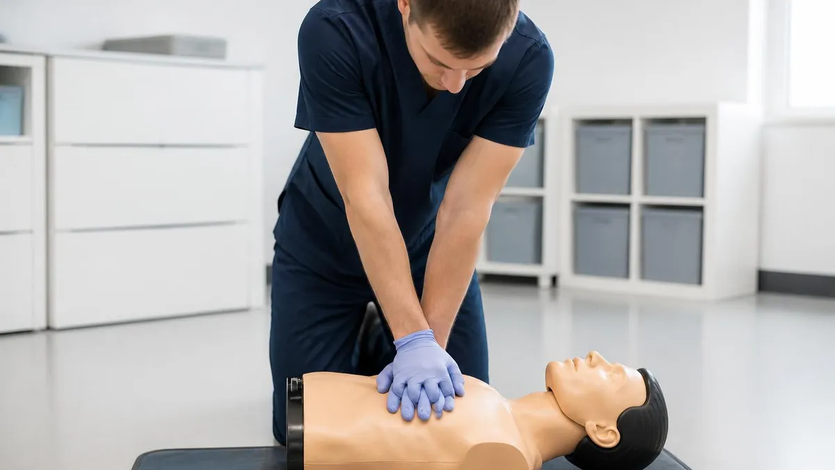
Free Online CPR Certification: Complete Guide to Legitimate Courses, Costs, and Workplace Acceptance in 2026
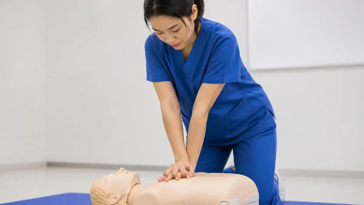
CPR Instructor Certification 2026: Complete Path to Teaching CPR

CPR AED Training Classes: Complete Guide to Certification, Courses, and Life-Saving Skills
How this page was made: written with AI tools and published by Nathan Beja, founder of Practice Test Geeks, who is responsible for its content. Exam facts are checked against the official sources listed below. Not reviewed by a subject-matter expert. Editorial policy
Sources & references
Exam facts on this page (fees, format, dates, passing scores) are checked against these official sources; the date is our last check. Editorial policy
- redcross.org — Score validity, renewal, test delivery, passing score · verified Oct 2026
- elearning.heart.org — Fees, score validity, passing score · verified Oct 2026
- shopcpr.heart.org — Test delivery · verified Oct 2026