Difference of Anatomy and Physiology: A Complete Guide to Understanding Structure vs. Function
Learn the difference of anatomy and physiology — how structure and function define the human body. Clear explanations, examples, and practice tests.

The difference of anatomy and physiology is one of the most foundational concepts in the biological and health sciences, yet it trips up students and curious learners alike every year. Put simply, anatomy is the study of structure — what body parts exist, where they are located, and how they relate spatially to one another — while physiology is the study of function — how those structures operate, communicate, and sustain life. Understanding this distinction from the very start sets the stage for everything else you will encounter in a health sciences curriculum.
Think of it this way: if the human body were a high-performance automobile, anatomy would be the engineering blueprint that labels every bolt, sensor, and panel, while physiology would be the owner's manual explaining how pressing the accelerator sends fuel to the pistons and ultimately moves the wheels. Both disciplines are indispensable, but they ask fundamentally different questions. Anatomy asks, "What is here and where is it?" Physiology asks, "What does it do and how does it do it?"
Historically, anatomy developed first, largely because it could be observed directly through dissection. Ancient physicians like Galen and, later, Andreas Vesalius meticulously described bones, muscles, and organs centuries before scientists had the tools to understand cellular metabolism or nerve conduction. Physiology emerged as a rigorous science during the 17th and 18th centuries, when William Harvey's landmark description of blood circulation demonstrated that structure and function are deeply intertwined disciplines that must be studied together.
In modern health education — nursing, physical therapy, medical school, athletic training, and allied health programs — these two subjects are almost always taught in the same course sequence precisely because they reinforce each other so powerfully. You cannot truly understand why the left ventricle has thicker walls than the right ventricle without knowing that the left side must generate enough pressure to push blood all the way to the toes, a physiological demand that shapes anatomical structure over evolutionary time and individual development.
Students preparing for credentialing exams, college entrance tests, or professional certifications consistently rank anatomy and physiology among the most challenging subjects they encounter. The sheer volume of terminology — with Latin and Greek roots describing thousands of structures and processes — can feel overwhelming. Building a solid mental framework around the core distinction between structure and function provides an anchor point from which all other knowledge can grow, making study sessions more efficient and retention far more durable.
This guide walks you through every major dimension of the anatomy-physiology distinction: the precise definitions, the overlapping subdisciplines, real-world clinical examples, and practical strategies for mastering both subjects on your own schedule. Whether you are a high school student taking your first biology elective, a college sophomore in a pre-nursing program, or a working professional brushing up for a continuing education exam, you will find clear explanations and actionable advice throughout every section below.
By the time you finish reading, you will be able to explain the difference confidently, recognize it in clinical scenarios, and apply that knowledge directly to your practice tests and coursework. Let's start by looking at some hard numbers that illustrate just how significant these disciplines are in the real world of healthcare education.
Anatomy and Physiology by the Numbers

Core Definitions: Anatomy vs. Physiology
The scientific discipline that examines the structure of living organisms — their shape, size, location, and spatial relationships. Anatomy ranges from gross (visible to the naked eye) to microscopic levels including histology and cytology.
The scientific discipline that investigates how living organisms and their parts carry out the chemical and physical functions of life, from cellular metabolism to whole-body responses like temperature regulation and blood pressure control.
The combined study recognizing that structure and function are inseparable. Understanding why a structure looks the way it does requires knowing what it must accomplish physiologically, and vice versa.
Both disciplines operate across the same hierarchy: chemical → cellular → tissue → organ → organ system → organism. Anatomy describes what exists at each level; physiology explains what happens there.
The overlap between anatomy and physiology is not accidental — it is built into the nature of living systems. Every anatomical structure has a physiological purpose, and every physiological process depends on a precise anatomical arrangement. The form-follows-function principle, a cornerstone of biological science, states that the shape and composition of any body structure reflect the tasks that structure must perform. Nowhere is this more apparent than in the cardiovascular system, where the heart's four chambers, thick muscular walls, and one-way valves are perfectly engineered for the rhythmic pumping that oxygenates every cell in your body.
Consider the alveoli of the lungs. Anatomically, they are tiny, balloon-like air sacs clustered at the ends of the bronchiolar tree, each surrounded by a dense capillary network and lined with an extraordinarily thin epithelial layer just one cell thick. Why this particular structure? Because physiology demands it: gas exchange — the movement of oxygen into the blood and carbon dioxide out — requires both maximum surface area and minimum diffusion distance. The anatomical features exist precisely to serve the physiological need, and studying one without the other leaves a gaping hole in understanding.
The nervous system offers another vivid illustration. Anatomically, a neuron consists of a cell body, dendrites, an axon, and synaptic terminals. Some axons are coated with a fatty myelin sheath. Physiologically, myelin dramatically increases the speed of electrical signal conduction — from roughly 1 meter per second in unmyelinated fibers to over 100 meters per second in fully myelinated ones. Diseases like multiple sclerosis, which destroy the myelin sheath, directly demonstrate what happens when anatomy is compromised: physiological function — motor control, sensation, cognition — breaks down in parallel.
In the skeletal system, the microstructure of bone illustrates the same principle on a cellular scale. Compact bone is organized into cylindrical units called osteons, each centered on a Haversian canal carrying blood vessels and nerves. The concentric rings of mineralized matrix provide enormous compressive strength while remaining surprisingly lightweight. This architectural choice is not arbitrary; it is the anatomical answer to the physiological problem of supporting body weight and resisting mechanical stress during movement without adding excessive mass that would slow the organism down.
Muscle tissue further blurs the line between the two disciplines. Skeletal muscle fibers contain parallel bundles of actin and myosin filaments — the anatomical scaffolding — that slide past each other during contraction, the physiological event. The ratio of slow-twitch (Type I) to fast-twitch (Type II) fibers in a given muscle reflects its primary function: postural muscles holding you upright all day are rich in fatigue-resistant Type I fibers, while sprint muscles need the explosive power of Type II fibers. Anatomy encodes physiological history and evolutionary pressure in every fiber arrangement.
Endocrine physiology and anatomy interweave elegantly in the pancreas, which contains both exocrine tissue (acinar cells secreting digestive enzymes into ducts) and endocrine tissue (the islets of Langerhans releasing insulin and glucagon directly into the bloodstream). Knowing that these two functionally distinct cell populations are anatomically intermingled within the same gland helps explain why pancreatic diseases — from pancreatitis to diabetes — can simultaneously affect digestion and blood sugar regulation in the same patient.
For students, recognizing this deep integration means that memorizing anatomical structures in isolation is a poor study strategy. Instead, linking each structure to its function — asking "why does this look this way?" every time you encounter a new term — creates richer memory traces, improves problem-solving on clinical scenario questions, and builds the kind of flexible knowledge that transfers from textbook to exam to real-world practice in healthcare settings across the United States.
Key Branches and Subdisciplines of Anatomy and Physiology
Gross anatomy (also called macroscopic anatomy) is the study of structures large enough to be seen with the naked eye, typically through cadaveric dissection, medical imaging, or surface observation. Regional anatomy organizes the body by area — head and neck, thorax, abdomen — while systemic anatomy groups structures by organ system across the entire body. Surface anatomy focuses on external landmarks used by clinicians to locate underlying structures, such as palpating the femoral pulse or identifying vertebral levels by counting spinous processes along the spine.
Microscopic anatomy dives below the threshold of unaided vision. Histology examines tissue types — epithelial, connective, muscular, and nervous — using stained slide preparations under light or electron microscopes. Cytology focuses at the individual cell level, studying organelle structure and membrane characteristics. Developmental anatomy (embryology) traces how structures form from fertilization through birth, explaining why certain congenital anomalies occur and what embryological layer each organ system originates from, knowledge that is critical for understanding malformation syndromes encountered in pediatric and obstetric clinical settings.

Studying Anatomy vs. Physiology: What to Expect
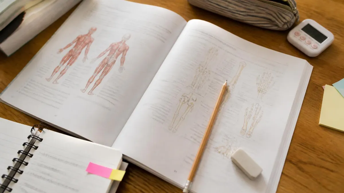
- +Anatomy rewards visual learners — diagrams, models, and color-coded atlases make structure intuitive and memorable
- +Anatomical knowledge is highly stable over time — the femur's basic structure has not changed, so mastered content stays useful throughout your career
- +Strong anatomy foundation makes clinical procedures (injections, catheter placement, surgical assisting) immediately safer and more accurate
- +Anatomy terminology follows systematic Latin and Greek roots, so learning root words multiplies vocabulary efficiently across hundreds of terms
- +Dissection and imaging labs provide tactile, hands-on learning experiences that cement spatial understanding in ways lectures alone cannot
- +Anatomy courses build transferable skills in precise observation and careful, methodical description applicable to many scientific fields
- −The sheer volume of memorization — over 10,000 named structures in the human body — can be cognitively overwhelming without a clear system
- −Without physiological context, anatomy can feel like rote memorization of names without understanding why any structure matters clinically
- −Cadaveric lab access is not always available in online or hybrid programs, limiting the hands-on experience that best consolidates spatial learning
- −Anatomy knowledge without physiology can give false confidence — knowing where the heart is does not explain how cardiac output is regulated
- −Regional vs. systemic approaches can create confusion when students switch between textbooks or course formats mid-curriculum
- −Clinical anatomy courses (for medical school) go significantly deeper than allied health versions, and students who switch programs may find major gaps
Anatomy and Physiology Mastery Study Checklist
- ✓Define anatomy and physiology in your own words without referencing notes to confirm baseline understanding
- ✓Identify at least one example from each of the 11 organ systems where structure directly enables function
- ✓Learn the six levels of structural organization from chemical to organism level and be able to place any term correctly
- ✓Practice using directional terms (superior, inferior, medial, lateral, proximal, distal) until they feel automatic in all body positions
- ✓Use a coloring atlas or digital 3D model to trace the cardiovascular circuit from heart through systemic and pulmonary loops
- ✓Write a one-sentence physiology explanation for every major anatomical structure you memorize to reinforce integration
- ✓Create a chart comparing the four tissue types — epithelial, connective, muscular, nervous — by structure and primary function
- ✓Complete at least two full-length practice tests under timed conditions before any scheduled exam to assess pacing and weak areas
- ✓Review all incorrect practice test answers by returning to the relevant anatomical structure and its physiological role
- ✓Use spaced repetition flashcard software (Anki or similar) with image-based cards for structures and process-based cards for mechanisms
Form Always Follows Function — Use This to Decode Any A&P Question
When an exam question describes an unfamiliar structure, ask yourself what physiological demands it must meet based on its described features. Thick walls suggest high pressure or strength demands. Large surface area suggests exchange or absorption. Dense vascularity suggests high metabolic activity. This reasoning strategy eliminates wrong answers on multiple-choice questions even when you cannot recall the exact term being described.
Clinical applications of the anatomy-physiology distinction are everywhere in healthcare, and understanding them deeply is what separates a competent health professional from an exceptional one. Take hypertension as an example. The physiological problem is elevated blood pressure — excessive force exerted by blood against arterial walls.
But the anatomical consequences are what make hypertension so dangerous over time: chronic high pressure causes the left ventricular wall to hypertrophy (thicken), the arterial walls to stiffen through a process called arteriosclerosis, and the kidneys' glomerular capillaries to sustain microscopic damage that gradually reduces filtration function. Anatomy and physiology spiral together in every chronic disease.
In orthopedics, understanding the difference proves essential for accurate diagnosis. A patient reports knee pain. The physiological complaint is pain and reduced range of motion. But determining the cause requires anatomical precision: is the pain arising from the medial meniscus, the anterior cruciate ligament, the patellar tendon, the infrapatellar bursa, or the articular cartilage of the femoral condyle? Each of these structures occupies a specific anatomical location, and each has a distinct physiological role. Palpation, special orthopedic tests, and imaging are all tools for mapping a physiological symptom back to a specific anatomical source.
Pharmacology and nursing provide equally rich examples. When a nurse administers an intramuscular injection into the ventrogluteal site, that choice reflects anatomical knowledge (the site avoids the sciatic nerve and superior gluteal vessels) and physiological knowledge (muscle tissue is highly vascularized, enabling rapid drug absorption compared to subcutaneous fat). Choosing the wrong site risks nerve damage — an anatomical catastrophe — while choosing the right site exploits physiological advantages of skeletal muscle perfusion. Every clinical skill integrates both disciplines simultaneously.
Respiratory therapy offers a stunning window into real-time anatomy-physiology integration. During mechanical ventilation, a respiratory therapist sets tidal volume based on the patient's lung anatomy (estimated by ideal body weight and height, which predict lung size) and adjusts inspiratory pressure and PEEP (positive end-expiratory pressure) based on physiological measurements of lung compliance, airway resistance, and oxygenation status. Getting these parameters wrong causes ventilator-induced lung injury — a case where insufficient physiological knowledge leads to anatomical harm at the alveolar level.
Neurology provides perhaps the most dramatic clinical example. Stroke — interruption of blood flow to a brain region — produces predictable neurological deficits based entirely on which anatomical territory is affected and what physiological functions that area subserves. Occlusion of the left middle cerebral artery affects Broca's area and the motor cortex controlling the right hand and face, producing expressive aphasia and right-sided weakness. A clinician who knows both the anatomy of cerebrovascular territories and the physiology of cortical functions can localize a stroke from bedside examination alone, before any imaging is performed.
Physical therapists apply the same integrated reasoning to rehabilitation. After a total knee replacement, a PT must understand the anatomy of the replaced joint — the implant's positioning relative to the femur, tibia, and patella — alongside the physiology of wound healing, the timeline of collagen remodeling, and the neuromotor re-education required to restore proprioception. Too much stress too soon (a physiological misjudgment) risks loosening the implant (an anatomical consequence). Too little activity delays muscle re-strengthening and prolongs disability. Clinical expertise lies in threading that needle correctly using knowledge from both disciplines simultaneously.
For students preparing for licensure exams across all of these healthcare fields, the message is consistent: you cannot afford to compartmentalize anatomy and physiology into separate mental silos. Every NCLEX-RN question about cardiovascular assessment, every NPTE question about biomechanics, every NBCOT question about upper extremity function draws simultaneously on structural knowledge and functional understanding. Building the habit of always asking both "what is it?" and "what does it do?" is the single most effective cognitive strategy for exam success and professional competency.
Many students use "anatomy" and "physiology" as if they mean the same thing, but conflating them leads to imprecise answers on exams and dangerous gaps in clinical reasoning. An anatomy question asks about location, structure, or relationship; a physiology question asks about mechanism, process, or regulation. When exam questions use these terms specifically, your answer must match the question's level — describing where a valve is located does not explain how it prevents blood backflow.
Preparing for any exam that covers anatomy and physiology requires a strategy that honors both the breadth of the content and the depth of integrated understanding the test demands. The first step is always an honest self-assessment: sit down with a blank sheet of paper and try to sketch the major organ systems from memory, labeling key structures and writing their primary functions beside each label. The gaps that emerge — the systems you struggled to recall, the functions you could not articulate — become the roadmap for your study plan over the following weeks and months.
Time management matters enormously in A&P preparation. Most successful students divide their study time roughly 60/40 between new material acquisition and active recall practice. The 60 percent acquisition phase uses textbooks, video lectures, digital atlases, and anatomical models to build initial understanding.
The 40 percent recall phase uses flashcards, practice tests, blank diagrams to label, and self-explanation exercises where you narrate a physiological process aloud as if teaching it to someone else. The teaching-out-loud technique is particularly powerful because it forces you to identify the exact moment your understanding becomes vague — and that moment marks where you need to return to the source material.
Spaced repetition is the gold standard for vocabulary-heavy content like anatomical terminology. Rather than studying all 50 muscle names for the upper limb in a single three-hour session, distribute that review across five sessions separated by 24 to 48 hours. Each time you successfully recall a term, increase the interval before the next review. Each time you fail to recall, reset the interval to one day. This approach exploits the spacing effect, a robust finding in cognitive science that dramatically increases long-term retention compared to massed practice sessions that cram all review into one block.
Practice tests deserve special attention as a study tool rather than just an assessment tool. When you answer an anatomy or physiology question incorrectly, resist the urge to simply read the correct answer and move on. Instead, trace the reasoning backward: what anatomical structure was being referenced, what is its physiological role, and what feature of that structure or function made the correct answer correct and your chosen answer wrong? This three-step post-error analysis builds the error-correcting metacognitive skills that separate students who plateau at 70 percent accuracy from those who push into the 90s.
Group study can accelerate learning when structured properly. The most effective group sessions assign each participant a topic to teach to the group, then have the group ask challenging questions. This peer-teaching format forces active retrieval, exposes hidden misunderstandings, and generates the kind of elaborative discussion — "wait, but how does that relate to what we learned about the autonomic nervous system?" — that weaves isolated facts into an interconnected web of knowledge. Passive group study, where everyone reads in silence and occasionally compares notes, provides far less benefit per hour invested.
Understanding the difference of anatomy and physiology also means knowing when each type of thinking is called for during an exam. Anatomy questions often use image-based formats showing diagrams, microscope slides, or imaging studies. Physiology questions more often use scenario-based formats describing a patient with specific symptoms or lab values and asking you to identify the underlying mechanism. Practicing both question types is essential — a student who only practices text-based physiology questions may freeze when encountering a histology slide identification item, even if they understand the underlying concepts perfectly well.
Finally, connect your study to real-world clinical contexts as often as possible. Read brief case studies, watch clinical procedure videos, and if your program offers simulation lab time, prioritize it. When anatomical and physiological knowledge is tied to a memorable clinical story — "the patient had crushing chest pain because atherosclerotic plaque ruptured in the left anterior descending artery, blocking oxygen delivery to the anterior myocardium" — it becomes dramatically harder to forget than a decontextualized list of terms and definitions. Context is the most powerful memory enhancer available to a health sciences student.
Practical tips for day-to-day anatomy and physiology learning can make the difference between grinding through material and actually enjoying the process. Start every new body system with a five-minute "big picture" overview before diving into details: what is this system's primary job, what are its main components, and how does it interact with the two or three systems most closely related to it? This orienting step prevents the disorientation that comes from memorizing the names of 15 kidney structures without any sense of why the kidney exists or what problem it solves for the organism.
Use multiple representational formats for each major concept. For the action potential, for example: read the textbook description of sodium and potassium ion movements; watch a 5-minute animated video showing the membrane voltage graph in real time; draw the graph yourself from memory; explain the process aloud using the analogy of a row of dominoes falling sequentially; then answer five multiple-choice questions testing different aspects of the mechanism. Engaging with the same concept through five different modes produces far deeper encoding than reading the same passage five times.
Body system integration exercises are particularly valuable for physiology. Pick two systems — say, the respiratory and cardiovascular systems — and spend 20 minutes mapping every physiological interaction you can identify: how does a drop in blood oxygen tension signal the carotid bodies, which relay to the respiratory center in the medulla, which increases breathing rate, which raises alveolar oxygen tension, which enables greater hemoglobin saturation, which delivers more oxygen to cardiac muscle, which improves cardiac output? Tracing these feedback loops builds the multi-system reasoning that exams increasingly demand.
Vocabulary acquisition benefits from etymology. The word "cardiovascular" combines the Greek "kardia" (heart) and Latin "vasculum" (small vessel). The word "pulmonary" derives from the Latin "pulmo" (lung). "Hepatic" comes from the Greek "hepar" (liver). Once you internalize 30 to 40 common roots, prefixes, and suffixes from Greek and Latin, you can decode unfamiliar anatomy and physiology terms you encounter for the first time on an exam — an invaluable skill when question writers introduce uncommon terms to test whether you can reason under pressure rather than simply recall memorized lists.
Sleep and exercise are not optional components of an effective A&P study plan — they are physiological requirements for memory consolidation. During slow-wave sleep, the hippocampus replays the day's learning and transfers it to cortical long-term storage. This is why all-night cramming sessions before anatomy practical exams consistently underperform compared to studying for a reasonable number of hours and then sleeping a full night.
Aerobic exercise has been shown in multiple peer-reviewed studies to increase BDNF (brain-derived neurotrophic factor), a protein that promotes synaptic plasticity and supports the formation of new memories. A 30-minute walk on study days is not procrastination — it is neurologically sound preparation.
When you feel overwhelmed by the volume of content, return to the core organizing principle of this guide: anatomy is structure, physiology is function, and they are inseparable. Every time you learn a new term, add a functional tag to it. Every time you encounter a physiological process, ground it in an anatomical location. This bidirectional linking strategy transforms an intimidating mountain of isolated facts into an interconnected, navigable knowledge network that grows stronger with each new piece of information you add to it.
Practice tests on PracticeTestGeeks.com are designed specifically to build this integrated thinking. Rather than testing anatomy and physiology in isolation, the questions place both types of knowledge in realistic clinical and scientific scenarios that mirror the format and difficulty level of professional licensing and certification exams. Regular practice under timed conditions, combined with careful error analysis after each session, is the fastest proven path from confusion to confidence in anatomy and physiology — no matter which credential or career you are working toward.
Anatomy Physiology Questions and Answers

Anatomy and Physiology Definitions: The Complete Beginner's Guide to Key Terms and Concepts

Anatomy and Physiology for Dummies: The Ultimate Beginner's Guide to Understanding the Human Body

Define Anatomy and Physiology: A Complete Guide to Understanding Human Body Structure and Function

Anatomy and Physiology Coloring Workbook: The Complete Guide to Visual Learning, Active Recall, and Mastering Body Systems

Difference Between Physiology and Anatomy: A Complete Guide to Understanding Structure vs. Function
About the Author
Educational Psychologist & Academic Test Preparation Expert
Columbia University Teachers CollegeDr. Lisa Patel holds a Doctorate in Education from Columbia University Teachers College and has spent 17 years researching standardized test design and academic assessment. She has developed preparation programs for SAT, ACT, GRE, LSAT, UCAT, and numerous professional licensing exams, helping students of all backgrounds achieve their target scores.