
Ch 4 Anatomy and Physiology: The Complete Student Guide to Mastering Chapter 4
Master ch 4 anatomy and physiology with this complete guide. Key concepts, study tips, and practice tests. 🎯 Everything students need.

When students reach ch 4 anatomy and physiology, they often encounter one of the most conceptually rich and demanding sections of their entire course. Chapter 4 typically covers tissue types, cellular organization, and the fundamental building blocks that connect individual cells to complete organ systems.
Understanding this material is not just about passing a test — it is the foundation upon which every subsequent chapter builds. Without a firm grasp of tissues, epithelial membranes, and connective tissue classifications, students struggle to make sense of how organs actually function. This guide walks you through everything you need to know to master the material.
Chapter 4 in most standard anatomy and physiology textbooks — including Marieb, Saladin, and OpenStax — centers on the concept of tissue organization. The human body contains four primary tissue types: epithelial, connective, muscle, and nervous. Each category has its own structural characteristics, locations throughout the body, and specific physiological roles. Epithelial tissue covers body surfaces and lines cavities. Connective tissue supports and binds other tissues together. Muscle tissue enables movement. Nervous tissue transmits electrical signals. Recognizing these categories and their subtypes is the core skill tested in chapter 4 assessments.
One of the most challenging aspects students face in this chapter is memorizing the extensive classification system for connective tissue. Unlike epithelial tissue, which is classified primarily by cell shape and layer count, connective tissue has a sprawling taxonomy that includes loose connective tissue, dense connective tissue, cartilage, bone, and blood. Each subtype has distinctive extracellular matrix components, cell types, and physiological functions. Students who invest time in understanding the logic behind these classifications — rather than rote memorization — consistently perform better on exams and retain the knowledge longer for clinical applications.
Epithelial tissue classification is another pillar of chapter 4 content. Epithelium is categorized both by the number of cell layers (simple versus stratified) and by cell shape (squamous, cuboidal, or columnar). Pseudostratified columnar epithelium and transitional epithelium add additional nuance to this system. Understanding where each type is found in the body — for example, simple squamous epithelium lining blood vessels versus stratified squamous epithelium covering the skin — gives the classification system meaningful context that aids long-term retention. You can explore chapter 4 anatomy and physiology difficulty in detail to set realistic expectations for this section.
Membranes are another major topic within chapter 4. Cutaneous, mucous, serous, and synovial membranes each represent a distinct tissue combination with unique structural and functional properties. The cutaneous membrane is the skin itself, the body's largest organ. Mucous membranes line body cavities that open to the outside world. Serous membranes line closed internal cavities and secrete lubricating fluid. Synovial membranes line joint cavities. These distinctions show up repeatedly on quizzes and exams, and clinical programs including nursing, physical therapy, and physician assistant training treat this knowledge as a prerequisite for everything from pharmacology to surgical technique.
Tissue repair and wound healing also appear in chapter 4 and represent a physiologically fascinating area that connects anatomy to real clinical scenarios. When tissues are damaged, the body initiates a predictable sequence of events: inflammation, organization, and restoration or fibrosis depending on the tissue type. Epithelial and connective tissues generally regenerate more efficiently than muscle or nervous tissue, which is why spinal cord injuries and heart attacks carry such serious long-term consequences. Understanding this regeneration gradient helps students appreciate why anatomy has direct implications for medical prognosis and treatment planning in healthcare careers.
Whether you are a nursing student, pre-med, exercise science major, or taking anatomy and physiology as a general education requirement, mastering chapter 4 will pay dividends throughout your academic program. The tissue types introduced here will reappear in every subsequent chapter covering organ systems. Students who can quickly identify tissue types from microscopic images, recall the distinguishing features of each connective tissue subtype, and explain membrane classifications are the ones who find later chapters significantly more manageable. Use the study tools and structured breakdowns throughout this guide to build the strong foundation chapter 4 demands.
Chapter 4 Anatomy and Physiology by the Numbers
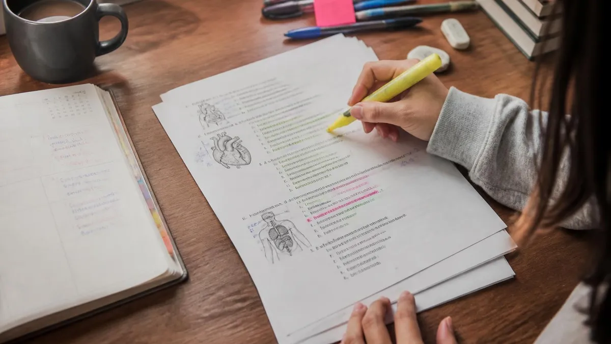
Core Topics Covered in Chapter 4
Learn to categorize epithelial tissue by cell shape (squamous, cuboidal, columnar) and layer count (simple, stratified, pseudostratified). Know where each type is located in the body and its primary physiological function, from gas exchange to secretion.
Master the diverse connective tissue family: loose areolar, dense regular, dense irregular, reticular, adipose, cartilage (hyaline, fibrocartilage, elastic), bone, and blood. Each type has unique matrix composition, cell types, and structural roles in the body.
Chapter 4 introduces the three muscle tissue subtypes — skeletal, cardiac, and smooth — and the basics of nervous tissue including neurons and neuroglia. These are previewed here and explored in greater depth in later chapters on muscular and nervous systems.
Understand the four membrane types: cutaneous (skin), mucous (lines cavities open to outside), serous (lines closed cavities), and synovial (lines joint spaces). Each has distinct tissue composition and function, and all appear frequently on anatomy quizzes and nursing exams.
Learn the stages of tissue repair — inflammation, organization, and regeneration or fibrosis — and understand why some tissues (epithelial, connective) heal well while others (cardiac muscle, nervous tissue) have limited regenerative capacity after injury.
Epithelial tissue is typically the first major category students encounter in chapter 4, and for good reason: it covers more surface area than any other tissue type and performs an extraordinary range of functions depending on its location. Simple squamous epithelium, composed of flat cells arranged in a single layer, is optimized for diffusion and filtration. You find it lining the alveoli of the lungs, where oxygen must move rapidly across into the bloodstream, and in the capillary walls where fluid and nutrients exchange between the blood and tissues. Its thinness is its defining functional feature.
As you move up the complexity ladder, cuboidal and columnar epithelium take on more active roles. Simple cuboidal epithelium lines kidney tubules and many glands, where it handles secretion and absorption with equal efficiency. Simple columnar epithelium lines most of the digestive tract, and its cells often bear microvilli to maximize surface area for nutrient absorption. Goblet cells interspersed among columnar cells secrete protective mucus. These structural features are not arbitrary — every detail of epithelial cell morphology reflects a specific functional demand placed on that tissue by its location in the body.
Stratified epithelia serve protective functions. Stratified squamous epithelium, the most common stratified type, forms the outer layer of skin (keratinized) and lines the mouth, esophagus, and vagina (non-keratinized). The multiple cell layers act as a physical barrier against abrasion, pathogens, and dehydration. As cells are lost from the surface through normal wear, the basal layer continuously divides to replace them. This regenerative strategy is uniquely suited to surfaces that endure constant mechanical stress, making stratified squamous epithelium a recurring topic in both chapter 4 and the integumentary system chapter that typically follows.
Connective tissue represents the most structurally diverse category in chapter 4. Unlike epithelial tissue, whose cells are packed tightly together, connective tissue cells are scattered within an extracellular matrix that the cells themselves produce. This matrix consists of ground substance — a gel-like material made of proteoglycans and glycoproteins — and protein fibers including collagen, elastic, and reticular fibers.
The ratio of matrix components to cells, and the specific fiber types present, defines each connective tissue subtype. Hyaline cartilage, for example, has abundant collagen but no visible fibers under standard histological staining, while fibrocartilage is dense with collagen bundles visible to the naked eye.
Adipose tissue deserves special attention in chapter 4 because students often underestimate its physiological importance. Far from being simply a passive energy reservoir, adipose tissue is now recognized as an endocrine organ that secretes hormones including leptin, adiponectin, and resistin — all of which influence metabolism, appetite regulation, and inflammatory responses. Brown adipose tissue, found mainly in newborns and around major blood vessels in adults, generates heat through a process called non-shivering thermogenesis. Understanding adipose tissue biology bridges chapter 4 material to the endocrine system and metabolic physiology covered later in most anatomy and physiology courses.
Blood is technically classified as a connective tissue in chapter 4, a fact that surprises many students initially. Blood meets the definition because it consists of cells (red blood cells, white blood cells, platelets) suspended in a liquid extracellular matrix called plasma. Like other connective tissues, blood develops from mesenchyme — embryonic connective tissue — and performs connective functions including transport, immune defense, and hemostasis. Recognizing blood as a connective tissue solidifies the conceptual framework for the entire connective tissue classification system and helps students see the underlying logic that organizes the category.
Nervous tissue, though introduced only briefly in chapter 4 in most curricula, establishes the conceptual vocabulary students will need for the nervous system chapters ahead. Neurons are the signaling cells, characterized by a cell body, dendrites that receive signals, and a long axon that transmits them.
Supporting cells called neuroglia — including astrocytes, oligodendrocytes, microglia, and Schwann cells — outnumber neurons roughly ten to one and perform essential supportive, protective, and metabolic functions. Grasping the basic neuron anatomy now makes the action potential, synaptic transmission, and neural pathway content in later chapters far less daunting for students who invest time in chapter 4 thoroughly.
Study Strategies for Chapter 4 Anatomy and Physiology
Creating a tissue classification matrix is one of the most effective memorization strategies for chapter 4. Draw a table with tissue types as rows and key features — cell shape, layers, location, function — as columns. Fill it in from memory, then check your textbook. Repeating this exercise three times over 48 hours leverages spaced repetition, which research consistently shows produces stronger long-term retention than a single intensive study session of equivalent total time.
Mnemonic devices can dramatically speed up memorization of connective tissue subtypes. For example, remember the three cartilage types as "Hyenas Fight Elastically" for Hyaline, Fibrocartilage, and Elastic cartilage. Similarly, grouping epithelial types by function — protection (stratified), absorption (simple columnar), diffusion (simple squamous), secretion (simple cuboidal) — gives each tissue type a functional identity that is far stickier than location-based memorization alone.

Advantages and Challenges of Chapter 4 as a Foundation Chapter
- +Establishes the universal vocabulary used in every subsequent organ system chapter
- +Tissue classification logic is internally consistent, making patterns learnable rather than purely memorized
- +Clinical relevance is immediately apparent — tissues connect directly to disease, injury, and treatment
- +Histology slide skills developed in chapter 4 are directly transferable to later lab practicals
- +Understanding tissue repair biology provides insight into why some injuries heal and others do not
- +Chapter 4 concepts are revisited and reinforced throughout the entire course, strengthening retention
- −The connective tissue taxonomy is extensive and requires systematic study over multiple sessions
- −Epithelial tissue subtypes with similar names (pseudostratified, stratified) are frequently confused on exams
- −Histology slide identification requires lab access or high-quality online resources not always available
- −Students without a strong biology background may find cell biology prerequisites lacking
- −Chapter 4 material can feel abstract until later chapters provide clinical application context
- −The sheer volume of vocabulary terms creates a significant memorization burden early in the course
Chapter 4 Anatomy and Physiology Study Checklist
- ✓Identify all four primary tissue types and state their general functions from memory
- ✓Classify epithelial tissues correctly by cell shape and number of layers for all six types
- ✓Name the three subtypes of cartilage and describe where each is found in the body
- ✓Distinguish loose areolar, dense regular, and dense irregular connective tissue by fiber arrangement
- ✓Explain why blood is classified as a connective tissue despite its liquid matrix
- ✓Describe the four body membrane types and provide one anatomical example of each
- ✓Outline the three stages of tissue repair and identify which tissue types regenerate versus scar
- ✓Practice identifying at least six tissue types from unlabeled histology slide images
- ✓Create a comparison table contrasting skeletal, cardiac, and smooth muscle tissue features
- ✓Complete at least two full practice tests covering chapter 4 content before your scheduled exam
Structure Always Reflects Function in Tissue Biology
Every structural feature of every tissue type in chapter 4 exists because it serves a specific physiological function. When you ask yourself WHY a tissue looks the way it does — why simple squamous epithelium is flat, why dense regular connective tissue has parallel fiber bundles — you stop memorizing and start understanding. This functional reasoning approach cuts study time significantly and produces answers you can reconstruct on exams even if you forget the exact terminology.
Preparing effectively for chapter 4 exams requires understanding the specific question formats your instructor uses. Multiple-choice questions in anatomy and physiology typically fall into three categories: recall questions that test whether you know a definition or classification, application questions that present a scenario and ask you to identify the relevant tissue type, and analysis questions that ask you to explain why a tissue is suited to its environment. Each category demands a different preparation strategy, and students who practice all three formats perform significantly better than those who focus exclusively on flashcard-style recall.
Image identification questions deserve special preparation time because many students ignore them until the night before an exam. Your instructor may show you a microscope slide image and ask you to name the tissue, or show you a labeled diagram and ask about the function of a highlighted cell type.
The best preparation strategy is to gather 20 to 30 unlabeled histology images from an online atlas — searching for each tissue name in your course glossary — and practice identifying them until you can name every one correctly without hesitation. This process typically requires two to three focused practice sessions of 45 minutes each.
Time management during chapter 4 exams is a skill in itself. Anatomy and physiology tests often cover a large number of items in a constrained time window, which means students who spend too long on difficult questions risk running out of time for questions they actually know. Develop a pacing strategy before exam day: decide in advance how many minutes you will allocate per question, set a mental checkpoint at the halfway point of the exam, and practice answering questions under time pressure during your study sessions so the exam environment does not feel unfamiliar or anxiety-provoking.
Essay and short-answer questions on chapter 4 material commonly ask students to compare and contrast tissue types, explain the relationship between tissue structure and function, or describe the tissue repair process. These questions reward students who have internalized the conceptual frameworks rather than memorized isolated facts. When writing an essay answer, start with a clear classification statement, provide the structural key features, connect those features to function, and give a specific anatomical example. This four-part structure consistently earns full marks and demonstrates the depth of understanding instructors are looking for in upper-level anatomy responses.
Laboratory practicals add a hands-on dimension to chapter 4 assessment that many students find more challenging than the written exam. In a practical, you may be given a microscope slide or anatomical model and asked to identify structures, label cell types, or describe tissue function within a strict time limit. Preparation for practicals requires direct experience with the materials — you cannot substitute textbook reading for time spent actually looking through a microscope at prepared slides. Ask your instructor or lab coordinator about open lab hours where you can practice independently before the scheduled practical exam date.
Online practice tests are among the most efficient tools available for chapter 4 exam preparation because they provide immediate feedback and expose you to question formats you might not encounter in your textbook review questions. Look for practice tests that include tissue identification images, not just text-based questions.
Many students find that their weakest area is not content knowledge but rather image interpretation — they know what hyaline cartilage is, but cannot identify it from a microscope slide. Targeted practice on image-based questions in the weeks before your exam is the fastest way to close this gap and convert borderline passing scores into strong grades on chapter 4 assessments.
Study groups work particularly well for chapter 4 content because the material lends itself to collaborative quiz formats. One student describes a tissue scenario — for example, "a tissue with no blood supply, cells in lacunae, and a glassy matrix" — while others race to identify it. This format builds both recall speed and conceptual reasoning simultaneously.
Students who have taught a concept to a peer consistently report higher confidence and retention than those who studied the same material alone, a phenomenon known in educational psychology as the protégé effect that is especially powerful for complex classification systems like those in chapter 4.

Many students lose significant points on chapter 4 exams by confusing pseudostratified columnar epithelium with stratified columnar epithelium — they look similar but are fundamentally different in structure and location. Pseudostratified appears layered but is actually a single cell layer where nuclei sit at different heights. True stratified columnar is genuinely multiple layers. Similarly, hyaline cartilage and fibrocartilage are frequently confused — remember that fibrocartilage has visible collagen bundles under a microscope, while hyaline has a smooth, glassy matrix with no visible fibers.
The knowledge you build in chapter 4 anatomy and physiology does not stay locked inside the classroom — it follows you directly into healthcare and science careers. Nurses use tissue biology knowledge daily when assessing wound healing, explaining skin conditions to patients, or understanding why certain medications target specific tissue types. A nurse who understands the difference between a first-degree burn affecting only the epithelial layer and a third-degree burn destroying all layers including subcutaneous connective tissue can communicate prognosis and treatment rationale far more accurately than one who learned anatomy by rote memorization alone.
Physical therapists and occupational therapists draw on chapter 4 concepts when designing rehabilitation programs. Understanding that dense regular connective tissue — tendons and ligaments — heals slowly due to poor vascularity informs decisions about how aggressively to rehabilitate a patient after a ligament repair. Knowing that skeletal muscle has limited regenerative capacity compared to epithelial tissue shapes expectations about recovery timelines and helps therapists set realistic goals with patients. This practical application of tissue biology makes chapter 4 far more than an academic exercise for students entering rehabilitation sciences.
Medical laboratory scientists and histotechnologists make tissue identification the centerpiece of their professional work. Pathologists rely on histological analysis — the microscopic examination of tissue sections — to diagnose cancers, inflammatory conditions, and infectious diseases. Chapter 4 provides the normal tissue baseline that makes pathological changes detectable. A histotechnologist who does not deeply understand normal epithelial tissue architecture cannot recognize the cellular disorganization that characterizes early carcinoma in situ. This diagnostic chain from tissue anatomy to clinical pathology makes chapter 4 one of the highest-value chapters in the entire anatomy and physiology curriculum for pre-health students.
Pharmacology is another field where chapter 4 tissue knowledge pays dividends. Drug delivery routes — oral, intravenous, transdermal, inhalation — each exploit the properties of specific tissue types. Transdermal patches rely on the permeability characteristics of the stratum corneum, the outermost keratinized layer of stratified squamous epithelium. Inhaled medications take advantage of the enormous surface area and thin simple squamous epithelium of the alveoli.
Oral medications must survive the enzymatic environment of the digestive tract's simple columnar epithelium before absorption. Understanding these routes at the tissue level builds the mechanistic thinking that distinguishes excellent pharmacology students from those who memorize routes without understanding the underlying biology.
Exercise physiology and sports medicine professionals apply chapter 4 knowledge to performance optimization and injury prevention. Hyaline cartilage covers the articulating surfaces of bones in synovial joints, and its avascular nature means that cartilage damage heals poorly without surgical intervention. Understanding this limitation informs athlete screening programs, equipment design, and training load management decisions.
The synovial membrane that lines joint cavities — a specialized connective tissue — secretes synovial fluid whose viscosity and composition can be altered by training, aging, and inflammatory disease. Sports medicine professionals who understand joint tissue biology at this level provide more effective care than those who treat the joint as a black box.
Even non-clinical career paths benefit from a strong chapter 4 foundation. Science writers, health educators, medical illustrators, and public health professionals all communicate tissue biology to non-specialist audiences. A health educator explaining why smoking damages the respiratory epithelium needs to understand simple columnar and pseudostratified columnar epithelium well enough to translate that knowledge accurately and accessibly.
A medical illustrator creating diagrams of wound healing must understand the sequence of tissue repair stages to represent them accurately. The conceptual depth that comes from genuine chapter 4 mastery supports professional excellence across a far wider range of careers than most students realize when they first open their textbooks to this chapter.
For students who want to understand just how demanding the broader course is before diving deeper into chapter 4, our guide on chapter 4 anatomy and physiology difficulty levels provides an honest assessment of what to expect across all course chapters. Building a strong tissue biology foundation now will make every subsequent topic — from the cardiovascular system to the endocrine system — more manageable and more meaningful throughout your anatomy and physiology studies.
Practical study tips for chapter 4 begin with breaking the content into manageable daily chunks rather than attempting to absorb all four tissue types in a single marathon session. Dedicate one focused study session to epithelial tissue classification and location, a second session to connective tissue taxonomy including all subtypes, a third to muscle and nervous tissue basics plus membrane types, and a fourth to tissue repair and integration review. This four-session structure distributes the cognitive load evenly and allows each session to end with a sense of accomplishment rather than overwhelm from the sheer volume of chapter 4 material.
Color-coding your notes is a surprisingly powerful organizational technique for chapter 4 content. Assign one color to epithelial tissue, another to connective tissue, a third to muscle tissue, and a fourth to nervous tissue. When you review your notes, the color coding allows you to quickly locate related information and reinforces the categorical boundaries between tissue types. Students who use consistent color-coding systems in their anatomy notes report faster review times before exams and fewer organizational errors on classification questions where the tissue type itself is the answer.
Drawing tissue diagrams from memory — without reference to your textbook — is one of the highest-yield study activities available for chapter 4. For each tissue type, attempt to sketch the cell arrangement, note the presence or absence of extracellular matrix, indicate the fiber types where relevant, and label the key cell types. Compare your drawing to the textbook illustration after completing it. The discrepancies you find reveal exactly which structural details you have not yet internalized, giving you a precise target for your next study session that is far more efficient than re-reading entire textbook sections.
Using multiple learning modalities for chapter 4 consistently outperforms single-modality approaches. Students who read the textbook, watch a video lecture, draw diagrams, complete practice questions, and discuss concepts with peers retain information across all those encoding channels simultaneously. When one pathway is blocked — for example, if you cannot recall a tissue name — another pathway (visual memory of the diagram, auditory memory of the instructor's voice) often provides an alternative retrieval route. Building this multi-modal memory network is especially important for chapter 4 because the exam will access your knowledge through multiple question formats including text, images, and diagrams.
Sleep is an underrated study tool that is especially important when learning complex classification systems like those in chapter 4. Research in cognitive neuroscience consistently shows that memory consolidation — the process of transferring information from short-term to long-term memory — occurs primarily during sleep.
Students who study chapter 4 material in the evening and then get seven to eight hours of sleep retain significantly more the following morning than students who stay up late cramming additional information at the cost of sleep duration. Building a study schedule that ends each session two to three hours before your planned bedtime gives your brain time to wind down and enter the consolidation phase effectively.
Practice testing yourself under exam conditions — closed book, timed, no notes — at least once before your actual chapter 4 exam. Many students avoid this because it feels uncomfortable to confront what they do not know, but this discomfort is precisely what makes simulated testing so valuable.
The retrieval effort required to answer a question without notes strengthens the memory far more than re-reading the correct answer passively. Aim to complete at least 40 practice questions covering all chapter 4 subtopics under timed conditions, review every incorrect answer thoroughly, and repeat the exercise 24 to 48 hours later to confirm the corrections have consolidated into accessible long-term memory.
Finally, connect chapter 4 content to clinical cases whenever possible to make the abstract concrete. When you learn about simple squamous epithelium lining the alveoli, think about what pneumonia does to those thin cells and why impaired gas exchange causes the breathing difficulty associated with that infection.
When you learn about hyaline cartilage covering joint surfaces, think about osteoarthritis as the gradual erosion of that cartilage and why joint replacement surgery has become one of the most common orthopedic procedures. These clinical anchors transform chapter 4 from a list of tissue classifications into a dynamic biological narrative that is both memorable and professionally meaningful for healthcare-bound students.
Anatomy Physiology Questions and Answers

How Hard Is Anatomy and Physiology? The Complete Honest Guide for Students

Eye Anatomy and Physiology: The Complete Guide to Understanding How Your Eyes Work
Anatomy and Physiology PDF: Complete Free Download Guide for 2026 Students
Human Anatomy and Physiology Book: The Complete Guide to Choosing, Using, and Mastering the Best Textbooks for 2026

Best Way to Learn Anatomy and Physiology: A Complete Student Guide
About the Author

Educational Psychologist & Academic Test Preparation Expert
Columbia University Teachers CollegeDr. Lisa Patel holds a Doctorate in Education from Columbia University Teachers College and has spent 17 years researching standardized test design and academic assessment. She has developed preparation programs for SAT, ACT, GRE, LSAT, UCAT, and numerous professional licensing exams, helping students of all backgrounds achieve their target scores.