Anatomy and Physiology Coloring Workbook Answer Key: Complete Chapter-by-Chapter Study Guide
Master anatomy with the coloring workbook answer key chapter 9 and beyond. 📚 Step-by-step guidance, study tips, and free practice tests inside.

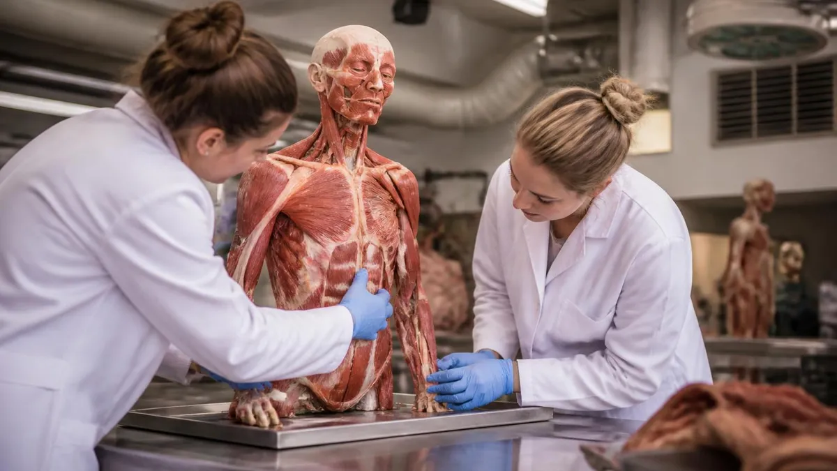
The anatomy and physiology coloring workbook answer key chapter 9 is one of the most searched resources among nursing students, pre-med undergraduates, and allied health learners across the United States. Chapter 9 typically covers the muscular system, and the coloring exercises inside Elaine Marieb's workbook are designed to help students internalize complex muscle names, origins, insertions, and actions through active engagement.
Unfortunately, without a verified answer key, students often spend hours second-guessing their colored diagrams, unsure whether they've correctly identified the sternocleidomastoid from the scalenes or the biceps brachii from the brachialis. This guide will walk you through every major chapter, with special focus on chapter 9, so you can study with confidence and accuracy.
The Anatomy and Physiology Coloring Workbook, authored by Elaine N. Marieb, has been a classroom staple since the 1980s. It accompanies her flagship textbook and is widely assigned in community colleges, four-year universities, and vocational nursing programs. The workbook is praised for transforming passive reading into active learning — a technique supported by cognitive science research showing that motor activity (coloring, labeling) encodes visual information more deeply than re-reading alone. Knowing the correct answers before you begin reinforces the feedback loop that makes this method work so powerfully.
Many students feel lost when they reach chapter 9 because the muscular system introduces hundreds of new Latin and Greek terms simultaneously. Muscles are named by location (brachialis), shape (deltoid), origin and insertion (sternocleidomastoid), number of heads (biceps, triceps), fiber direction (transverse abdominis), and function (flexor carpi radialis). Keeping all of these naming conventions organized is a genuine cognitive challenge, and mistakes in the workbook — left uncorrected — can harden into exam-day misconceptions. Having access to a reliable anatomy and physiology coloring workbook answer key is therefore not cheating; it is smart, error-correcting study practice.
Chapter 9 in most editions of the Marieb workbook opens with an overview of muscle tissue types before narrowing to skeletal muscle structure. Students label the sarcomere, identify the A band, I band, H zone, and Z disc, then move on to whole-muscle anatomy including fascia, epimysium, perimysium, and endomysium. Each coloring plate builds on the last, so a mistake in plate one can cascade through the entire chapter. Working from the answer key lets you verify each plate before advancing, keeping your conceptual foundation solid as complexity increases.
Beyond chapter 9, the workbook covers eleven major body systems across roughly 350 pages and dozens of coloring plates. Topics range from the skeletal system (chapters 5–7) and nervous system (chapters 11–13) to the endocrine, cardiovascular, respiratory, digestive, urinary, and reproductive systems. Students who use the workbook systematically — completing every plate, checking answers, and reviewing errors — consistently report higher exam scores than peers who rely solely on reading the textbook. The coloring method forces you to slow down, make decisions, and engage with spatial relationships that text descriptions alone cannot capture.
This article provides chapter-by-chapter guidance for using the coloring workbook effectively, explains what the answer key covers in each major section, and offers study strategies that top anatomy students use to turn workbook sessions into lasting, exam-ready knowledge. Whether you are struggling with chapter 9's muscular system, confused about the cardiovascular plates in chapter 18, or simply looking for a more efficient way to prepare for your next lab practical, this comprehensive resource has you covered. Free practice quizzes linked throughout will help you test your knowledge as you go.
Understanding the structure of the workbook itself is the first step toward using it well. Each chapter begins with a brief orientation section, followed by labeling exercises, coloring plates, and short-answer review questions. The coloring plates always include a color key legend that tells you which structure should be assigned which color. When using the answer key, cross-reference both the color assignments and the label placements, because both dimensions carry exam-relevant information. Getting the color right but misplacing a label — or vice versa — still represents incomplete learning that the answer key helps you catch and correct immediately.
Anatomy Coloring Workbook by the Numbers

What Each Major Workbook Chapter Covers
Covers anatomical terminology, body planes, organ systems overview, the cell, and basic tissue types. Answer key plates focus on cell organelle labeling, epithelial tissue identification, and connective tissue classification.
Bone structure, the axial and appendicular skeleton, joints, and articulations. Students label bones, foramina, and joint types. Answer key ensures correct identification of 206 named bones and major surface features.
The most complex chapter pair in the workbook. Covers sarcomere structure, major muscle groups by region, origins, insertions, and actions. Chapter 9 answer key is critical for preventing cascading labeling errors.
Neurons, spinal cord cross-sections, brain regions, cranial nerves, autonomic nervous system, and special senses. Answer key plates include detailed brain labeling and reflex arc diagrams used heavily on practicals.
Covers endocrine, cardiovascular, lymphatic, respiratory, digestive, urinary, and reproductive systems. Each chapter uses multicolor plates to distinguish vessels, ducts, organs, and glands by functional category.
Chapter 9 of the Marieb coloring workbook is the chapter that separates committed anatomy students from casual ones. The muscular system introduces more new vocabulary per page than any other chapter in the book, and the coloring plates require students to differentiate between muscles that lie in close anatomical proximity.
The anterior thigh, for example, contains the rectus femoris, vastus lateralis, vastus medialis, and vastus intermedius — four distinct muscles that share a common distal tendon and are easy to conflate on a two-dimensional diagram. The chapter 9 answer key assigns different colors to each muscle and shows precisely where each label arrow terminates, eliminating the guesswork that trips up thousands of students each semester.
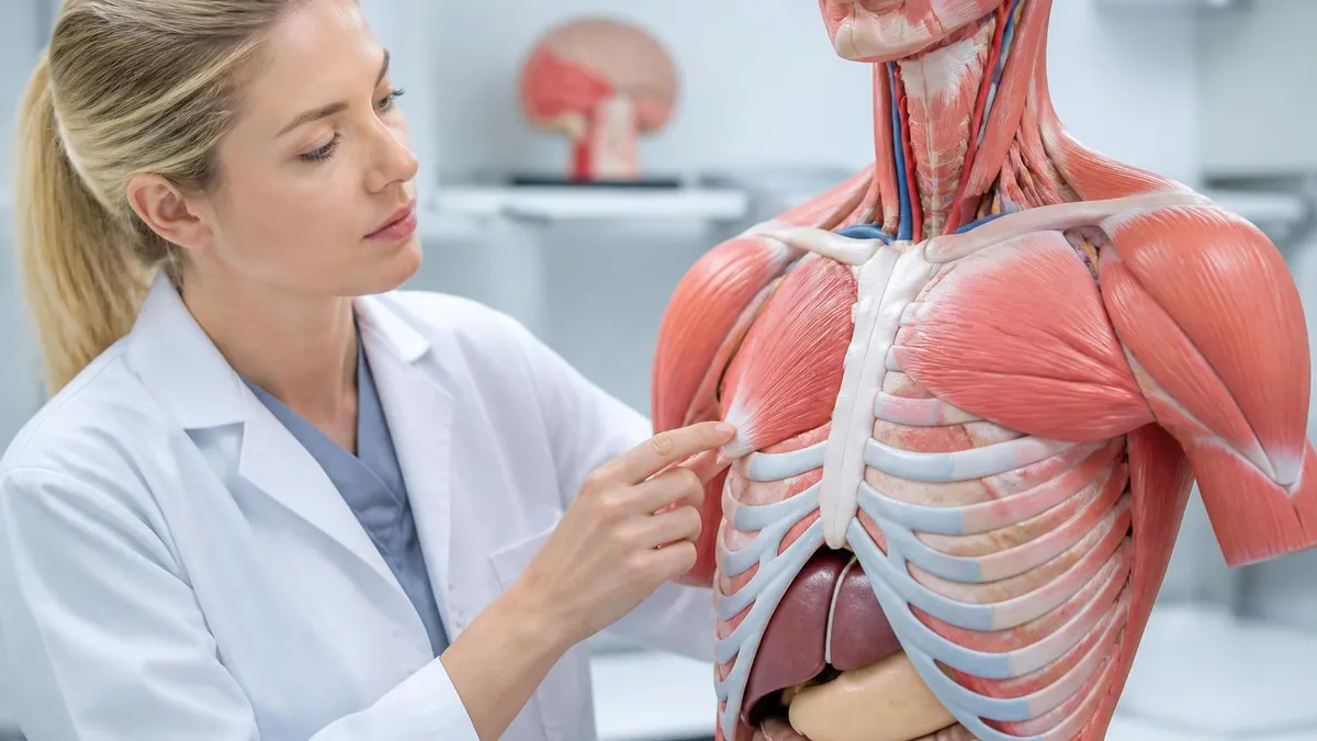
The chapter opens with microanatomy before moving to gross anatomy. The first several plates show the hierarchical organization of skeletal muscle: the whole muscle is wrapped in epimysium, bundles of fibers (fascicles) are wrapped in perimysium, and individual muscle fibers are wrapped in endomysium. Students then zoom in further to label the sarcomere — the functional unit of contraction — identifying the thick myosin filaments, thin actin filaments, titin elastic proteins, and regulatory proteins troponin and tropomyosin. Getting these sarcomere labels correct is foundational, because every subsequent discussion of muscle contraction in lecture will reference these structures by name.
After the microanatomy plates, chapter 9 moves through major muscle groups in a regional sequence. The head and neck muscles come first, including the masseter, temporalis, zygomaticus major, orbicularis oculi, and orbicularis oris. These muscles are frequently tested on lab practicals because students can observe and palpate them on their own faces, making misidentification particularly embarrassing.
The answer key for this section clarifies common confusions, such as the difference between the sternocleidomastoid (which originates on the sternum and clavicle and inserts on the mastoid process) and the scalene muscles (which originate on the cervical vertebrae and insert on the first two ribs).
The trunk muscles follow, and this is where many students experience their first major stumbling block. The abdominal wall has four distinct muscle layers whose fiber directions alternate to provide multidirectional strength: the external oblique runs inferiorly and medially (like hands in front pockets), the internal oblique runs superiorly and medially (perpendicular to external oblique), the transversus abdominis runs horizontally, and the rectus abdominis runs vertically.
Coloring each layer a distinct color while referencing the answer key helps you build an accurate three-dimensional mental model of the abdominal wall that will serve you throughout the rest of the course and in clinical practice.
Upper limb muscles in chapter 9 are organized by function: flexors and extensors of the elbow, rotators of the forearm, and the intrinsic muscles of the hand. Students frequently mix up the brachialis and biceps brachii because both flex the forearm, but the brachialis is the more powerful flexor, originating on the distal humerus rather than the coracoid process and supraglenoid tubercle.
The chapter 9 answer key color-codes these muscles distinctly and provides brief origin-insertion summaries in the margin that serve as built-in flashcards. If your edition does not include margin notes, supplement with index cards as you work through each plate.
Lower limb muscles are the final major section of chapter 9 and are among the most clinically relevant muscles in the body. The gluteal muscles (gluteus maximus, medius, and minimus), hip flexors (iliopsoas, rectus femoris, sartorius), hamstrings (biceps femoris, semitendinosus, semimembranosus), quadriceps, and leg muscles (gastrocnemius, soleus, tibialis anterior) all appear in this section. The answer key is especially helpful here because the posterior thigh and leg are dense with overlapping muscles that share similar names. Distinguishing the semitendinosus from the semimembranosus, or the peroneus longus from the peroneus brevis, requires precise labeling that the answer key provides.
Finally, chapter 9 concludes with a summary table of muscle actions organized by body region and movement type. Students are asked to match muscles to their prime mover, synergist, and antagonist roles for common movements like elbow flexion, knee extension, and shoulder abduction.
Completing this table while cross-referencing the answer key transforms isolated muscle facts into integrated functional knowledge — the kind of understanding that earns full credit on essay questions and impresses instructors during oral practicals. Spending an extra 30 minutes on this summary section during your first pass through the chapter will save hours of rote memorization later in the semester.
How to Use the Anatomy and Physiology Coloring Workbook Answer Key
Before you pick up a single colored pencil, read the entire chapter in your textbook and attend (or watch) the corresponding lecture. The coloring workbook is designed to reinforce material you have already encountered, not introduce it for the first time. Preview the plates in the workbook without filling them in, noting which structures appear most frequently and which relationships look most complex. Write a brief list of terms you are uncertain about — these will be your priority focus areas when you compare your work to the answer key.
Gather your materials: at minimum, 12 distinctly different colored pencils. Professional-grade pencils like Prismacolor allow better blending and more nuanced color distinction than cheap wax pencils. Label each pencil with a sticky note so you can quickly retrieve the same color for structures that span multiple plates. The answer key typically assigns the same color to the same structure type across the entire chapter, so consistency in your color selection reinforces learning through visual pattern recognition.

Coloring Workbook with Answer Key: Benefits and Limitations
- +Active coloring encodes spatial relationships better than passive reading or highlighting
- +Answer key provides immediate corrective feedback that prevents wrong information from solidifying
- +Color-coding creates visual memory hooks that surface during exams when you visualize your workbook pages
- +Systematic chapter-by-chapter structure mirrors most A&P course syllabi, making planning easy
- +Workbook exercises double as built-in practice questions with answer key verification
- +Completing every plate builds a personalized, annotated study guide you own through graduation
- −The workbook is designed for a specific Marieb textbook edition; answer key accuracy varies across editions
- −Coloring sessions are time-intensive — a complete chapter can take 3 to 5 hours of focused work
- −Two-dimensional diagrams cannot fully replicate the three-dimensional complexity of real cadaver anatomy
- −Students who copy the answer key without attempting plates first derive little cognitive benefit
- −Some answer keys available online contain errors that can reinforce incorrect information
- −Workbook alone is insufficient preparation for clinical lab practicals that use real specimens or 3D models
Chapter 9 Coloring Workbook Study Checklist
- ✓Read chapter 9 of the Marieb textbook completely before opening the workbook
- ✓Gather at least 12 distinctly different colored pencils and label them for consistency
- ✓Attempt each coloring plate independently before consulting the answer key
- ✓Mark every error with a red checkmark rather than erasing — errors are learning data
- ✓Identify the naming convention (location, shape, function) for every muscle you label
- ✓Complete the origin-insertion-action table for all major muscles in chapter 9
- ✓Sketch the anterior and posterior body outlines from memory after finishing the chapter
- ✓Score your blank sketches against the answer key and list your weakest structures
- ✓Schedule a second coloring session 48–72 hours after the first for spaced repetition
- ✓Take a free practice quiz to verify that workbook learning transfers to test-style questions
The 72-Hour Rule for Anatomy Retention
Educational psychology research consistently shows that reviewing anatomy material within 72 hours of initial learning reduces forgetting by up to 60%. Complete your chapter 9 coloring plates, then return to the same plates two to three days later without looking at your previous work. Students who apply this spaced-repetition principle to the Marieb coloring workbook report retaining muscle names, origins, and insertions through final exams — not just through the next weekly quiz.
One of the most common mistakes students make with the anatomy and physiology coloring workbook is treating it as a coloring book rather than a study tool. The goal is never beautiful artwork — the goal is accurate, error-corrected learning. Students who spend excessive time perfecting color gradients or decorative shading miss the educational point entirely.
Each plate should be completed quickly and accurately, with time invested in the verification and error-analysis steps rather than in aesthetic refinement. A plate finished in 20 minutes with careful answer-key comparison teaches more than a plate finished in 60 minutes of artistic coloring without verification.
Another frequent error is skipping the short-answer and fill-in-the-blank questions that appear between coloring plates. These questions test conceptual understanding rather than visual identification — they ask you to explain why the tibialis anterior is a prime mover for dorsiflexion, or to predict which muscles would be weakened by damage to the femoral nerve. Students who skip these sections gain visual memory without functional understanding, which becomes a serious liability on essay exams and clinical reasoning assessments in nursing and allied health programs.
Label arrows in the workbook plates can be ambiguous, particularly in densely populated regions like the forearm flexors or the deep muscles of the back. When an arrow could plausibly point to either of two adjacent structures, do not guess — consult your textbook's corresponding figure to see the regional anatomy in higher resolution.
The answer key will tell you which structure the arrow was intended for, but understanding why that arrow points there (not just that it does) requires the textbook cross-reference. Building this habit of cross-referencing saves you from rote memorization and builds the analytical skill that anatomy instructors prize most.
Students working on the cardiovascular system chapters (typically chapters 18–19 in the Marieb workbook) face a different challenge: the sheer number of named vessels. The arterial tree alone branches from the aorta through dozens of named arteries before reaching capillary beds, and the venous drainage follows a parallel but not always symmetrical path.
The answer key for these chapters is particularly valuable because it maintains consistent color coding — arteries in red, veins in blue — while still distinguishing individual vessels within each category through shade variation. Always replicate this convention in your own coloring to build the oxygenated/deoxygenated distinction that underlies cardiovascular physiology.
The nervous system chapters (11–15) present yet another challenge: the brain has no clean external landmarks that correspond to internal functional regions, making it genuinely difficult to know where the thalamus ends and the hypothalamus begins on a midsagittal section diagram. The answer key for these plates should be used not just to verify label placement but also to study the spatial relationships between structures.
How does the corpus callosum relate to the lateral ventricles? Where does the cerebellum sit in relation to the pons and medulla? These spatial relationships are constantly tested on both lab practicals and written exams, and the coloring workbook is one of the most effective tools for building accurate spatial mental models of brain anatomy.
For the endocrine, respiratory, and digestive system chapters, the answer key helps students master the distinction between anatomical structures and functional zones. The stomach, for example, has a cardia, fundus, body, and pylorus — regions with distinct histological and functional characteristics that must be correctly labeled. The small intestine's three segments (duodenum, jejunum, ileum) are distinguished by their mucosal features and lengths. The answer key directs students to use different colors for each region, reinforcing the functional distinctions through visual differentiation. This approach is far more effective than trying to memorize region boundaries from a list of numbers.
Finally, many students overlook the chapter review sections at the end of each workbook chapter, which typically include matching exercises, diagram completion activities, and case study questions. These sections are the closest the workbook comes to replicating exam-style assessment, and they should be completed under timed conditions before checking the answer key.
Treat each chapter review as a mini practice exam: set a timer, work through every question without referring to your completed plates, then grade yourself using the answer key. This performance data is your most accurate predictor of how prepared you are for the real exam, and it shows you exactly which sections of the chapter need another review pass before test day.

Not all anatomy and physiology coloring workbook answer keys available on the internet are accurate or edition-matched. The Marieb workbook has gone through 12 editions, and plate numbering, label placements, and even structure names have changed between editions. Always verify that any answer key you use matches your specific edition number (printed on the workbook's copyright page). Using a mismatched answer key can introduce errors that are worse than having no key at all, since incorrect information presented with apparent authority is harder to unlearn than a genuine gap in knowledge.
Developing a sustainable weekly study rhythm around the coloring workbook is the single most impactful thing you can do to succeed in anatomy and physiology. Most A&P courses move through one to two body systems every two to three weeks, which means students need to complete one to two workbook chapters per week to stay current.
Spreading this across three to four dedicated study sessions per week — rather than cramming all plates the night before an exam — dramatically improves retention and reduces pre-exam anxiety. Block your calendar for workbook sessions the same way you block time for class, treating them as non-negotiable academic commitments.
Study groups can amplify the benefits of the coloring workbook when used correctly. Rather than simply coloring together (which becomes social rather than educational), the most effective group format involves each member completing their own plates independently, then gathering to compare answers before consulting the key. Discrepancies between group members' answers reveal genuinely contested or ambiguous label placements that the instructor should be asked to clarify. This collaborative error-detection process catches mistakes that individual study misses and builds the collaborative clinical reasoning skills valued in healthcare professions.
Integrating the coloring workbook with other study resources multiplies its effectiveness. Pair each completed plate with a corresponding 3D anatomy app review (Complete Anatomy, BioDigital Human, or Visible Body are popular choices), a cadaver photograph from an atlas like Grant's or Netter's, and a short Khan Academy or professor-recorded video lecture. This multimodal approach ensures that workbook-derived knowledge transfers to three-dimensional real-world anatomy rather than remaining flat-image-dependent. Students who cross-reference across modalities consistently outperform those who rely on any single resource, including the workbook alone.
Flash card systems like Anki can be built directly from your coloring workbook sessions. As you verify each plate against the answer key, create one Anki card per structure: the front shows a blank version of the diagram with an arrow to the structure; the back shows the structure name, its origin, insertion, and primary action.
This converts your passive error-checking into active card creation, and the resulting Anki deck becomes a personalized, exam-calibrated review tool that you can use on your phone between classes. Students who maintain an Anki deck built from the coloring workbook report significantly higher lab practical scores than those who study only from the completed workbook pages.
Clinical correlation is another powerful technique that transforms coloring workbook sessions from academic exercises into professionally meaningful learning. As you complete each plate, ask yourself: where would I palpate this muscle on a patient? What movement would be lost if this nerve were damaged? What would a torn tendon at this insertion look like on an MRI?
These clinical questions are not just intellectually stimulating — they create the kind of meaningful, contextual memory encoding that survives the stress of clinical rotations and licensing exams years after your A&P course ends. Many nursing and physical therapy students report that their coloring workbook sessions were the most clinically useful hours of their undergraduate education.
Time management during exam week deserves special attention. In the 48 hours before a major anatomy exam, do not attempt to complete new coloring plates for the first time — the cognitive load of new plate completion is too high to manage alongside final review of already-learned material. Instead, use your completed plates as quick-review flash cards, covering the labels with a sheet of paper and testing yourself structure by structure.
Spend extra time on the structures you marked with red checkmarks during your original error-analysis sessions, as these are statistically your most likely exam vulnerabilities. This targeted review approach is far more efficient than re-reading the textbook or re-watching lecture videos at the last minute.
If you are a visual-spatial learner — someone who thinks in images and maps rather than words and lists — the anatomy and physiology coloring workbook is particularly well-suited to your learning style, and investing extra time in it relative to other study methods will pay disproportionate dividends.
If you are a verbal or auditory learner, the workbook is still valuable but should be supplemented with verbal repetition (saying muscle names and actions aloud as you color) and discussion-based review sessions. The answer key serves every learning style equally well as a calibration tool, but the strategies you layer on top of it should be tailored to how your brain best encodes and retrieves information.
Preparing effectively for anatomy and physiology exams requires more than completing coloring plates — it requires converting visual memory into retrieval-ready knowledge that performs under exam conditions. The best anatomy students treat the coloring workbook as phase one of a three-phase study cycle: first, color and verify against the answer key; second, test retrieval through blank-sketch exercises and practice quizzes; third, simulate exam conditions with timed, closed-book assessments. Each phase serves a distinct cognitive purpose, and skipping any one of them leaves gaps that the other two cannot fill.
Practice tests are the most underutilized study tool in anatomy and physiology, despite overwhelming research evidence that testing yourself is more effective than any form of passive review. After completing each workbook chapter and verifying your plates against the answer key, immediately take a practice quiz covering the same body system.
Free online quizzes — including those linked throughout this article — present the same anatomical facts in multiple-choice format, which trains the recognition skills needed for written exams. Lab practical preparation requires a different mode: labeling photographs of cadaver specimens or anatomical models, which you can practice using free online atlas resources.
Mnemonics and memory devices can bridge the gap between workbook learning and exam performance for structures that resist plain memorization. The carpal bones, cranial nerves, and branches of the brachial plexus all have well-established mnemonic sequences that anatomy students have relied on for generations. When the coloring workbook presents these high-density regions, supplement your coloring session with the relevant mnemonic, then test yourself on both the mnemonic and the corresponding plate label within the same study session. The dual encoding — visual from the plate, verbal from the mnemonic — creates two independent retrieval pathways that dramatically reduce exam-day blanking.
Instructors frequently design lab practicals using model specimens, plastinated cadaver sections, or digital 3D platforms rather than the flat diagrams in the workbook. This means that workbook mastery is necessary but not sufficient for practical exam success. Bridge this gap by using 3D anatomy apps during the same study session as your workbook coloring.
For every plate you complete and verify, spend five minutes rotating the corresponding 3D model on your app, identifying the same structures in three dimensions. This paired practice ensures that the knowledge you build from the workbook generalizes to the three-dimensional presentations used in lab practicals and clinical settings.
Finally, do not underestimate the value of teaching anatomy to others as a study technique. Once you have completed a chapter's coloring plates and verified them against the answer key, try explaining the chapter to a study partner who has not yet worked through it. Teaching forces you to organize your knowledge into a logical explanatory sequence, which reveals hidden gaps in your own understanding.
If you cannot explain why the gastrocnemius is both a knee flexor and a plantar flexor — or why the biceps brachii is named for its two heads rather than its function — you do not yet understand the material well enough for an A or B on the exam. Use those gaps as your next day's study targets.
Building cumulative review into your weekly schedule prevents the most insidious form of anatomy forgetting: the slow erosion of early-chapter material as later chapters demand increasing cognitive bandwidth. Many students know the skeletal system perfectly at midterm but cannot name five bones by finals because they stopped reviewing skeletal content after moving on to muscles. Block 20 minutes of every study session for cumulative review of previous chapters using your completed workbook plates. This ongoing review keeps early material accessible and prevents the desperate, ineffective cramming of forgotten content in the days before a comprehensive final exam.
The anatomy and physiology coloring workbook, used systematically with a verified answer key and paired with practice quizzes, spaced repetition, and multimodal review, is one of the most powerful study tools available to A&P students at any level.
Students who commit to this integrated approach from week one of their course consistently earn higher exam scores, perform more confidently on lab practicals, and retain anatomical knowledge longer than peers who rely on passive reading and last-minute cramming. The investment of time and attention that the coloring workbook demands is exactly proportional to the depth of understanding it builds — and that understanding is the foundation of every healthcare career.
Anatomy Physiology Questions and Answers

How Hard Is Anatomy and Physiology? The Complete Honest Guide for Students
Human Anatomy and Physiology Book: The Complete Guide to Choosing, Using, and Mastering the Best Textbooks for 2026

Eye Anatomy and Physiology: The Complete Guide to Understanding How Your Eyes Work
Anatomy and Physiology PDF: Complete Free Download Guide for 2026 Students

Anatomy and Physiology for Dummies: The Ultimate Beginner's Guide to Understanding the Human Body
About the Author
Educational Psychologist & Academic Test Preparation Expert
Columbia University Teachers CollegeDr. Lisa Patel holds a Doctorate in Education from Columbia University Teachers College and has spent 17 years researching standardized test design and academic assessment. She has developed preparation programs for SAT, ACT, GRE, LSAT, UCAT, and numerous professional licensing exams, helping students of all backgrounds achieve their target scores.