
Abdomen Anatomy and Physiology: The Complete Guide to Structures, Functions, and Clinical Relevance
Master abdomen anatomy and physiology with this complete guide covering organs, regions, digestive functions, and clinical relevance. 🎯 Perfect for students.

Understanding abdomen anatomy and physiology is one of the most essential foundations in any healthcare or life sciences curriculum. The abdomen is far more than just your stomach — it is a complex, densely packed cavity housing over a dozen organs responsible for digestion, filtration, hormone production, and immune defense. Students studying for nursing boards, medical entrance exams, or allied health certifications will encounter abdominal anatomy repeatedly, and a solid grasp of this region separates high scorers from those who struggle with clinical application questions.
The abdominal cavity extends from the diaphragm superiorly down to the pelvic brim inferiorly, bounded anteriorly by the abdominal wall muscles and posteriorly by the vertebral column and back muscles. Within this space, organs are organized into two primary categories: those within the peritoneal cavity (intraperitoneal) and those lying behind the peritoneum (retroperitoneal). Understanding this distinction matters clinically because it determines how organs move, how pain radiates, and how surgeons access these structures during procedures.
Clinicians and students alike use two main systems to describe abdominal locations: the four-quadrant system and the nine-region system. The four-quadrant method divides the abdomen into right upper, left upper, right lower, and left lower quadrants using two perpendicular planes through the navel. The nine-region system further subdivides these into the epigastric, umbilical, hypogastric, left and right hypochondriac, left and right lumbar, and left and right iliac regions — each corresponding to specific underlying organs and clinical presentations.
Physiologically, the abdomen orchestrates an extraordinary range of functions simultaneously. The gastrointestinal tract breaks down food through mechanical and chemical digestion, absorbing nutrients across nearly 20 feet of small intestine. The liver processes absorbed nutrients, detoxifies the blood, produces bile, and synthesizes essential proteins. The pancreas serves a dual role as both an endocrine organ secreting insulin and glucagon into the bloodstream and an exocrine organ releasing digestive enzymes into the small intestine. Each of these functions is tightly regulated by neural and hormonal signals.
For students preparing for anatomy examinations, the abdominal region often appears in multiple question formats. Multiple-choice questions might ask about organ locations within specific quadrants. Clinical vignettes frequently describe abdominal pain and expect students to identify which organ is involved based on location and radiation pattern. Practical lab components may require identification of structures on cadaveric specimens or anatomical models. Knowing not just where structures are but why they are positioned there — and what physiological roles they serve — gives students a powerful advantage on all of these question types.
The abdominal wall itself is a multilayered structure with significant physiological importance. Moving from superficial to deep, it consists of skin, subcutaneous fat, three layers of flat muscles (external oblique, internal oblique, and transverse abdominis), the rectus abdominis running vertically in the midline, and the peritoneum lining the cavity. These muscle layers work in concert to support the spine, help breathing, increase intra-abdominal pressure during defecation and childbirth, and protect the vulnerable organs beneath. Weakness or defects in this wall can result in hernias, a common clinical finding requiring surgical correction.
This comprehensive guide walks you through every major structure, physiological process, and clinical correlation within the abdomen. Whether you are approaching your first anatomy course, reviewing for a high-stakes licensing exam, or simply trying to build a more complete clinical picture, the information presented here will give you both the conceptual framework and the detailed knowledge you need to succeed. Each section is designed to build on the last, creating a layered understanding that transforms memorized facts into functional, applied knowledge.
Abdominal Anatomy by the Numbers

The Nine Abdominal Regions: What Each Contains
Located centrally below the sternum, the epigastric region contains the stomach, part of the liver, the duodenum, and the pancreas head. Pain here commonly signals gastritis, peptic ulcers, or pancreatitis — conditions every clinician must recognize quickly.
The central region surrounding the navel houses the transverse colon, loops of small intestine, and portions of the duodenum. Diffuse umbilical pain often indicates early appendicitis before it migrates to the right iliac region, a classic clinical sequence.
Found in the lower midline, this region contains the urinary bladder, uterus in females, and sigmoid colon. Suprapubic pain or pressure frequently signals urinary tract infections, bladder distension, or gynecological pathology requiring prompt evaluation.
The iliac fossae lie on either side of the hypogastric region. The right iliac fossa is critically important as the location of the appendix and cecum. Left iliac pain often points to sigmoid colon pathology including diverticulitis or irritable bowel syndrome.
The hypochondriac regions flank the epigastric area and house the liver and gallbladder on the right, spleen and gastric fundus on the left. The lumbar regions contain the ascending and descending colon plus portions of the kidneys — retroperitoneal structures protected by the posterior abdominal wall.
The liver is the largest solid organ in the abdominal cavity and one of the most metabolically active organs in the entire body. Weighing approximately 1,500 grams in adults, it occupies primarily the right upper quadrant and extends into the epigastric region.
The liver performs over 500 distinct functions, including processing glucose and lipids absorbed from the intestines, synthesizing clotting factors and plasma proteins like albumin, conjugating and excreting bilirubin, and detoxifying drugs, alcohol, and metabolic waste products. Its dual blood supply — receiving nutrient-rich blood from the portal vein and oxygenated blood from the hepatic artery — reflects its central metabolic role.
The gallbladder sits in a fossa on the visceral surface of the liver's right lobe and stores bile produced by hepatocytes. When fatty foods enter the duodenum, cholecystokinin (CCK) is released from intestinal enteroendocrine cells, stimulating gallbladder contraction and bile release through the common bile duct. Bile emulsifies dietary fats, breaking large lipid droplets into smaller micelles that pancreatic lipase can efficiently break down. Gallstones, formed when bile becomes supersaturated with cholesterol, affect roughly 20 million Americans and represent one of the most common abdominal surgical diagnoses encountered in clinical practice.
The pancreas is a retroperitoneal organ stretching transversely across the posterior abdomen, with its head nestled in the C-shaped curve of the duodenum and its tail reaching toward the spleen. As an exocrine gland, it produces approximately 1.5 liters of pancreatic juice daily, containing bicarbonate to neutralize gastric acid entering the duodenum and enzymes including amylase, lipase, and proteases in their inactive zymogen forms. As an endocrine gland, the islets of Langerhans release insulin from beta cells when blood glucose rises, glucagon from alpha cells when glucose falls, and somatostatin from delta cells to fine-tune both hormones' secretion.
The spleen, positioned in the left upper quadrant under the diaphragm, performs critical immunological and hematological functions. It filters blood, removing aged or damaged red blood cells and recycling their iron. The white pulp of the spleen contains lymphoid tissue that initiates immune responses against blood-borne pathogens, while the red pulp serves as a reservoir for platelets and acts as an emergency blood store. Despite its important functions, humans can survive splenectomy — though with increased susceptibility to encapsulated bacteria — because the liver and lymph nodes can partially compensate for its filtration and immune roles.
The kidneys, while technically retroperitoneal structures, are anatomically associated with the abdomen and appear on virtually every abdominal anatomy examination. Each kidney measures approximately 10 centimeters in length and filters roughly 180 liters of blood per day, ultimately producing 1–2 liters of urine. The nephron — the functional unit of the kidney — performs filtration at the glomerulus, selective reabsorption along the tubular system, and secretion of waste products. The adrenal glands sit atop each kidney and secrete cortisol, aldosterone, and catecholamines, regulating stress responses, fluid balance, and blood pressure.
The stomach occupies the left upper quadrant and epigastric region, serving as a muscular reservoir capable of expanding to hold up to 4 liters of ingested food and liquid. Its wall contains four principal layers: the mucosa (which secretes gastric acid, pepsinogen, intrinsic factor, and mucus), the submucosa, the muscularis (with three rather than the usual two muscle layers, enabling the churning motion that creates chyme), and the serosa. The stomach's rugae — longitudinal folds that flatten as the organ fills — dramatically increase surface area for secretion and allow for significant distension without tearing the mucosal lining.
The small intestine — comprising the duodenum, jejunum, and ileum — is where the vast majority of nutrient digestion and absorption occurs. Villi (finger-like projections) and microvilli (forming the brush border) increase the mucosal surface area to approximately 250 square meters — roughly the size of a tennis court — maximizing the efficiency of absorption. Nutrients absorbed here enter either the portal venous system (carbohydrates, amino acids, water-soluble vitamins) or the lymphatic lacteals (long-chain fatty acids, fat-soluble vitamins). The ileocecal valve at the junction of the ileum and large intestine regulates the passage of contents and prevents backflow.
Digestive Physiology: From Ingestion to Absorption
Mechanical digestion begins in the mouth with mastication and continues as the stomach churns food into chyme through coordinated contractions of its three-layered muscularis. The pyloric sphincter controls the release of chyme into the duodenum in small, regulated boluses approximately every 20 seconds, ensuring the small intestine is not overwhelmed and digestive enzymes have adequate contact time with food particles. Segmentation contractions in the small intestine mix chyme with digestive secretions rather than propelling it forward.
Peristalsis — coordinated waves of smooth muscle contraction — propels food through the entire GI tract from esophagus to rectum. The enteric nervous system, often called the "second brain," contains over 100 million neurons embedded in the gut wall and independently coordinates motility, secretion, and blood flow without requiring input from the central nervous system. This explains why intestinal function can be partially preserved even after spinal cord injuries, and why emotional stress — which activates the autonomic nervous system — profoundly affects bowel habits in conditions like irritable bowel syndrome.
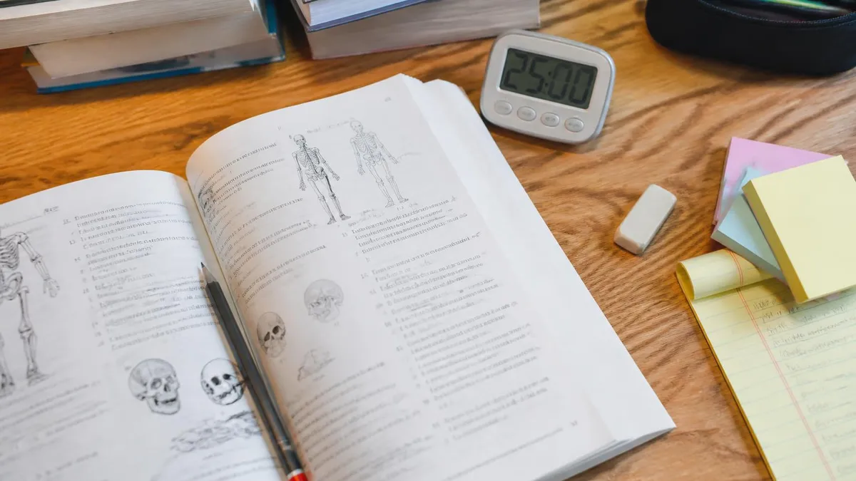
Studying Abdominal Anatomy: Visual Learning vs. Text-Based Study
- +Visual diagrams and labeled models accelerate spatial memory for organ locations and quadrant positions
- +Three-dimensional anatomical atlases reveal relationships between structures that flat diagrams cannot capture
- +Color-coded region maps help students distinguish overlapping organ boundaries quickly during timed exams
- +Interactive anatomy software allows virtual dissection, building procedural understanding alongside factual recall
- +Clinical imaging (CT scans, ultrasound) bridges the gap between textbook diagrams and real patient presentations
- +Drawing structures from memory reinforces recall pathways more effectively than passive re-reading of notes
- −Visual study without reading context can produce isolated facts that are hard to apply in clinical vignettes
- −Atlas diagrams often show idealized anatomy that differs from normal anatomical variation in real patients
- −Over-reliance on 2D diagrams poorly prepares students for 3D spatial questions on practical exams
- −Digital apps can become passive entertainment rather than active learning if not used with recall testing
- −Text-heavy physiology explanations without visual anchors make enzyme cascades and hormonal feedback loops difficult to retain
- −Students who skip the cellular and histological level often struggle with pathophysiology questions linking structure to disease
Pros and Cons at a Glance
| Pros | Cons |
|---|---|
| Visual diagrams and labeled models accelerate spatial memory for organ locations and quadrant positions | Visual study without reading context can produce isolated facts that are hard to apply in clinical vignettes |
| Three-dimensional anatomical atlases reveal relationships between structures that flat diagrams cannot capture | Atlas diagrams often show idealized anatomy that differs from normal anatomical variation in real patients |
| Color-coded region maps help students distinguish overlapping organ boundaries quickly during timed exams | Digital apps can become passive entertainment rather than active learning if not used with recall testing |
| Interactive anatomy software allows virtual dissection, building procedural understanding alongside factual recall | Text-heavy physiology explanations without visual anchors make enzyme cascades and hormonal feedback loops difficult to retain |
| Clinical imaging (CT scans, ultrasound) bridges the gap between textbook diagrams and real patient presentations | Students who skip the cellular and histological level often struggle with pathophysiology questions linking structure to disease |
Abdominal Anatomy Mastery Checklist for Exam Success
- ✓Identify all nine abdominal regions and list at least two organs or structures found in each
- ✓Draw the four-quadrant system and correctly place the liver, spleen, stomach, appendix, and bladder
- ✓Describe the difference between intraperitoneal and retroperitoneal organs with three examples of each
- ✓Explain the dual blood supply of the liver and why the portal vein is physiologically distinct from other veins
- ✓Trace the path of a fat molecule from ingestion through chylomicron formation to entry into the bloodstream
- ✓List the exocrine secretions of the pancreas, their targets, and the signals that trigger their release
- ✓Explain three physiological roles of the liver beyond bile production and give clinical examples of liver failure
- ✓Describe the layers of the abdominal wall from superficial to deep and the herniation risk at each weak point
- ✓Identify the three sections of the small intestine and the primary absorptive function unique to each region
- ✓Practice locating structures on an unlabeled diagram under time pressure to simulate exam conditions
McBurney's Point: Why One Landmark Matters for Every Student
McBurney's point — located one-third of the way from the right anterior superior iliac spine to the navel — marks the classic site of maximum tenderness in acute appendicitis. Every anatomy and physiology student should memorize this landmark because it appears frequently on nursing, medical, and allied health examinations. Rebound tenderness at this location (Blumberg's sign) suggests peritoneal irritation, signaling a surgical emergency. Understanding why this point matters requires knowing both the anatomical location of the appendix and the physiological consequence of its inflammation.
Abdominal pain is among the most common presenting complaints in emergency medicine, primary care, and surgical practices, making a thorough understanding of abdominal anatomy clinically indispensable. The pattern of pain — its location, radiation, character, and associated symptoms — provides critical diagnostic clues that depend entirely on which organ is affected and how its nerve supply maps onto the body surface. Visceral pain from hollow organs like the stomach, intestines, and gallbladder is transmitted through autonomic afferent fibers and is characteristically poorly localized, cramping or colicky in nature, and accompanied by nausea, sweating, and restlessness.
Somatic pain, in contrast, arises when disease or injury involves the parietal peritoneum, which is innervated by somatic sensory nerves with precise dermatome mapping. This is why a perforated appendix causes initially vague periumbilical pain (visceral phase from appendiceal distension) that later localizes sharply to the right lower quadrant (somatic phase once inflammation reaches the parietal peritoneum). This two-phase progression is a classic teaching point that appears in virtually every clinical anatomy curriculum and reflects the fundamental difference between visceral and somatic pain pathways.
Referred pain — sensation perceived at a site distant from the actual pathological process — further complicates abdominal clinical presentations. Gallbladder disease frequently refers pain to the right shoulder tip via the phrenic nerve (C3-C5), which shares the same dermatomal level as the diaphragm, which is irritated by bile or blood in the peritoneal space. Splenic rupture causes left shoulder pain by the same mechanism. Appendicitis can refer pain to the testicle in males because the appendix and gonadal structures share a common embryonic origin and thus a common nerve supply at the T10 spinal level.
The mesentery deserves special attention in clinical anatomy discussions because it serves far more functions than its historical description as a mere fatty tether for the small intestine. Modern anatomical research has reclassified the mesentery as a continuous organ extending from the duodenojejunal flexure to the upper rectum.
It contains the superior mesenteric artery and vein (supplying the entire small intestine and right colon), numerous lymph nodes critical for immune surveillance of gut-derived antigens, lymphatic channels for fat absorption, and neural plexuses that regulate gut motility. Mesenteric ischemia — inadequate blood flow through these vessels — causes one of the most catastrophic acute abdominal emergencies, with mortality rates exceeding 60% when diagnosis is delayed.
The peritoneum itself is a serous membrane that reduces friction between abdominal organs, confines infection and inflammation within specific compartments (a life-saving mechanism when the appendix ruptures), and serves as a potential space that can accumulate fluid (ascites) in liver disease, heart failure, or malignancy. The greater omentum — the large, apron-like fold of peritoneum hanging from the greater curvature of the stomach — acts as an immunological "policeman" of the abdomen, migrating toward sites of inflammation and infection to contain and combat pathological processes. Students who understand omental function can reason through otherwise puzzling clinical scenarios.
Abdominal aortic anatomy is equally critical for clinical correlation. The descending aorta enters the abdomen through the aortic hiatus of the diaphragm at T12 and branches successively into the celiac trunk (T12, supplying the foregut organs: stomach, liver, spleen, and proximal duodenum), the superior mesenteric artery (L1, midgut: distal duodenum through transverse colon), and the inferior mesenteric artery (L3, hindgut: descending colon through rectum). These arterial territories define embryological gut divisions that are reflected in surgical approach, transplant logistics, and the pattern of ischemic injury when vascular supply is compromised by atherosclerosis or thromboembolism.
For students preparing examinations on clinical anatomy, the abdominal vasculature consistently appears in both identification and application questions. The portal venous system — which drains the GI tract, spleen, and pancreas into the liver before blood reaches the systemic circulation — creates a clinically important pathway for both drug metabolism and pathological pressure elevation.
Portal hypertension, a consequence of liver cirrhosis, causes blood to be rerouted through collateral vessels including esophageal varices, rectal hemorrhoidal veins, and periumbilical vessels (caput medusae). Recognizing these collateral pathways on imaging or physical examination requires a thorough mental map of normal abdominal venous anatomy as the baseline for comparison.

Many students confuse the histological layers of different abdominal organs, particularly the GI wall layers (mucosa, submucosa, muscularis, serosa) with the peritoneal layers (parietal vs. visceral peritoneum) and the abdominal wall layers (external oblique, internal oblique, transverse abdominis). On high-stakes exams, questions frequently test whether you can correctly identify which layer is involved in specific pathologies — misidentifying a layer can lead to the wrong answer even when you understand the clinical concept correctly.
Mastering abdominal anatomy requires more than memorizing organ lists — it demands understanding the developmental logic behind why structures are positioned where they are. During embryological development, the primitive gut tube rotates 270 degrees counterclockwise (when viewed from the front) as it elongates and herniates temporarily into the umbilical cord before returning to the abdominal cavity.
This rotation explains why the duodenum ends up retroperitoneal and curved to the right, why the transverse colon passes in front of the small intestine, and why the cecum descends to the right lower quadrant rather than remaining in the upper abdomen where the primitive hindgut begins.
Malrotation of the gut — a developmental anomaly occurring in approximately 1 in 500 live births — results in abnormal positioning of intestinal structures and creates vulnerability to volvulus (twisting of the bowel on its mesentery), a life-threatening surgical emergency most common in neonates. Understanding normal rotation makes abnormal rotation immediately comprehensible — and explains why a midgut volvulus causes the characteristic double-bubble sign on imaging and must be corrected surgically within hours to prevent bowel necrosis. This is precisely the type of developmental-anatomical-clinical connection that distinguishes excellent anatomy students from those who have merely memorized structure names.
The large intestine — comprising the cecum, ascending colon, transverse colon, descending colon, sigmoid colon, and rectum — performs the final stages of digestive processing. Its primary functions are water and electrolyte absorption, compaction of feces, and housing the colonic microbiome.
The approximately 100 trillion bacteria inhabiting the large intestine — representing over 1,000 species — perform remarkable metabolic functions including synthesis of vitamin K and certain B vitamins, fermentation of indigestible dietary fiber into short-chain fatty acids that nourish colonocytes, and training of the mucosal immune system. The microbiome has emerged as a major focus of contemporary biomedical research, with alterations in its composition linked to obesity, inflammatory bowel disease, colorectal cancer, and even neuropsychiatric conditions.
The appendix, long dismissed as a vestigial structure with no physiological function, is now understood to serve as a "safe house" for beneficial gut bacteria, enabling re-inoculation of the colon following illness-related diarrhea. It also contains substantial lymphoid tissue (making it part of the gut-associated lymphoid tissue, or GALT) that contributes to mucosal immune responses, particularly in early life.
While appendectomy carries no significant long-term consequence for most adults, its removal in infancy may marginally affect the maturation of intestinal immune function — an active area of research that reflects how anatomical structures dismissed for decades are yielding new physiological insights with improved investigative tools.
Retroperitoneal structures deserve thorough attention because they are frequently tested and are anatomically distinct in ways that have clinical significance. In addition to the kidneys and adrenal glands, the retroperitoneum contains the inferior vena cava, the abdominal aorta, portions of the duodenum and pancreas, the ascending and descending colon, and major lymphatic vessels. Retroperitoneal bleeding — from trauma, ruptured aortic aneurysm, or pancreatic necrosis — is particularly dangerous because the retroperitoneal space can accommodate several liters of blood before clinical signs become apparent, leading to delayed diagnosis and higher mortality compared to intraperitoneal hemorrhage that manifests earlier with peritoneal signs.
The inguinal region — where the abdominal wall meets the thigh — contains the inguinal canal, a pathway through which the spermatic cord passes in males and the round ligament passes in females. The inguinal canal has anatomically weak points at the deep and superficial inguinal rings that predispose to herniation of abdominal contents.
Indirect inguinal hernias (entering through the deep inguinal ring, lateral to the inferior epigastric vessels) represent the most common type of abdominal hernia overall and are far more frequent in males due to incomplete obliteration of the processus vaginalis after testicular descent. Direct inguinal hernias protrude through Hesselbach's triangle, a region bounded medially by the rectus abdominis, inferiorly by the inguinal ligament, and laterally by the inferior epigastric vessels.
Femoral hernias — less common but more prone to incarceration — pass beneath the inguinal ligament through the femoral canal, which lies medial to the femoral vein. Because the femoral ring is rigid, contents that herniate through it are easily trapped and cut off from blood supply, making femoral hernias surgical emergencies far more frequently than inguinal hernias. Correctly distinguishing between inguinal and femoral hernias on physical examination requires precise anatomical knowledge of the relationship between the inguinal ligament, pubic tubercle, and femoral vessels — another example of how anatomical precision has direct, immediate clinical value that rewards thorough study.
If you want more targeted practice, the anatomy and physiology practice test page covers the same material with additional questions and explanations.
Effective exam preparation for abdominal anatomy begins with building a strong spatial foundation before layering in physiological details. Start by spending the first study sessions focused exclusively on location — draw blank outlines of the abdomen repeatedly and practice placing organs without reference materials. Research in educational psychology consistently shows that effortful retrieval practice (testing yourself rather than re-reading) produces dramatically superior long-term retention. Using flashcards for organ locations, setting daily self-quiz sessions, and drawing anatomical diagrams from memory are all forms of this high-yield retrieval practice that should anchor your study routine.
Once location is secure, shift focus to function. For each organ, ask three questions: what does it receive, what does it produce, and what regulates it? The liver receives portal blood containing absorbed nutrients and toxic byproducts; it produces bile, clotting factors, albumin, and glucose; it is regulated by insulin, glucagon, and the autonomic nervous system.
This functional framework turns a long list of organ facts into a coherent physiological story that is far easier to remember and apply. Students who study anatomy as a narrative — where each structure makes sense in the context of the whole system — consistently outperform those who approach it as a disconnected list of names and numbers.
Clinical correlation is the highest-yield strategy for locking anatomical knowledge into memory. For every structure you study, read about one clinical condition associated with it: gallstones for the gallbladder, pancreatitis for the pancreas, hepatic encephalopathy for the liver, Crohn's disease for the terminal ileum. When you later encounter that clinical condition on an exam, your brain automatically activates the associated anatomy — and vice versa. Medical and nursing educators recognize this bidirectional reinforcement, which is why clinical vignette questions dominate high-stakes licensing examinations rather than straightforward identification questions that reward pure memorization.
Practice questions are non-negotiable for exam success. No amount of reading or passive review substitutes for sitting down with timed questions that force you to retrieve, reason, and apply what you have studied. Work through questions covering anatomical identification, physiological mechanisms, clinical correlation, and developmental embryology.
Review every wrong answer in detail, identifying whether your error was factual (you did not know the information) or reasoning-based (you knew the facts but applied them incorrectly). These two types of errors require different remediation strategies: factual errors need more active retrieval practice, while reasoning errors need more deliberate practice with clinical vignettes that develop pattern recognition.
Group study and teaching are underutilized strategies for anatomy mastery. Teaching a concept to a peer forces you to organize your knowledge, identify gaps, and construct clear explanations — all cognitive processes that reinforce memory far more than solo reading.
Try teaching the entire digestive pathway from mouth to anus to a classmate, explaining at each step what happens mechanically, what enzymes are involved, what hormones are released, and what clinical problems can arise. If you can do this fluently and accurately, you have moved from surface-level familiarity to genuine mastery that will serve you through examination and into clinical practice.
Time management during anatomy examinations deserves deliberate practice. Many students spend disproportionate time on early questions and rush through later ones, resulting in avoidable errors on questions they would have answered correctly with more time. Practice taking full-length simulated exams under real time constraints, training yourself to mark uncertain questions, continue forward, and return efficiently. Abdominal anatomy sections often include images, diagrams, or imaging studies that take extra seconds to interpret — budget for this. Students who have drilled exam pacing alongside content knowledge almost uniformly outperform those who have studied content alone.
Finally, integrate your abdominal anatomy study with the broader systems it connects to. The abdomen does not function in isolation — it is deeply intertwined with the cardiovascular system (portal and systemic circulation), the endocrine system (pancreatic hormones, adrenal cortex), the immune system (lymphoid tissue, gut microbiome, GALT), and the nervous system (enteric, autonomic, and somatic innervation). Students who build these connections — rather than studying each organ system in a vacuum — develop the integrated, systems-level understanding that is increasingly emphasized on modern licensing examinations and that forms the true foundation of clinical reasoning.
Make connection-building a deliberate part of every study session, and your abdominal anatomy mastery will pay dividends across your entire medical or health science curriculum.
Sample Anatomy and Physiology Practice Questions
Try these questions from our free Anatomy and Physiology practice tests. The correct answer and an explanation follow each question.
The tricuspid valve is located between which two structures?
- A. Left atrium and left ventricle
- B. Right ventricle and pulmonary artery
- C. Right atrium and right ventricle
- D. Left ventricle and aorta
Answer: C. Right atrium and right ventricle
The tricuspid valve, named for its three cusps, separates the right atrium from the right ventricle and prevents backflow during contraction.
Which structure connects the left and right cerebral hemispheres?
- A. Corpus callosum
- B. Thalamus
- C. Hypothalamus
- D. Brainstem
Answer: A. Corpus callosum
The corpus callosum is a large band of white matter containing nerve fibers that connects and facilitates communication between the two cerebral hemispheres.
What is the function of the mitral (bicuspid) valve?
- A. Prevents backflow from the aorta into the left ventricle
- B. Prevents backflow from the left ventricle into the left atrium
- C. Prevents backflow from the pulmonary artery into the right ventricle
- D. Prevents backflow from the right ventricle into the right atrium
Answer: B. Prevents backflow from the left ventricle into the left atrium
The mitral valve sits between the left atrium and left ventricle, preventing backflow of blood into the atrium during ventricular contraction.
What is the main function of the lymphatic system?
- A. Transport oxygen in the blood
- B. Filter blood and remove waste
- C. Drain excess interstitial fluid and provide immunity
- D. Produce hormones for metabolism
Answer: C. Drain excess interstitial fluid and provide immunity
The lymphatic system collects and returns interstitial fluid to the blood, absorbs fats from the digestive tract, and is a critical part of the immune system, housing lymphocytes that defend against pathogens.
Anatomy Physiology Questions and Answers

How Hard Is Anatomy and Physiology? The Complete Honest Guide for Students

Eye Anatomy and Physiology: The Complete Guide to Understanding How Your Eyes Work
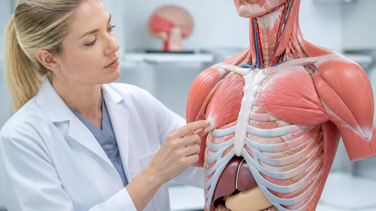
Anatomy and Physiology PDF: Complete Free Download Guide for 2026 Students
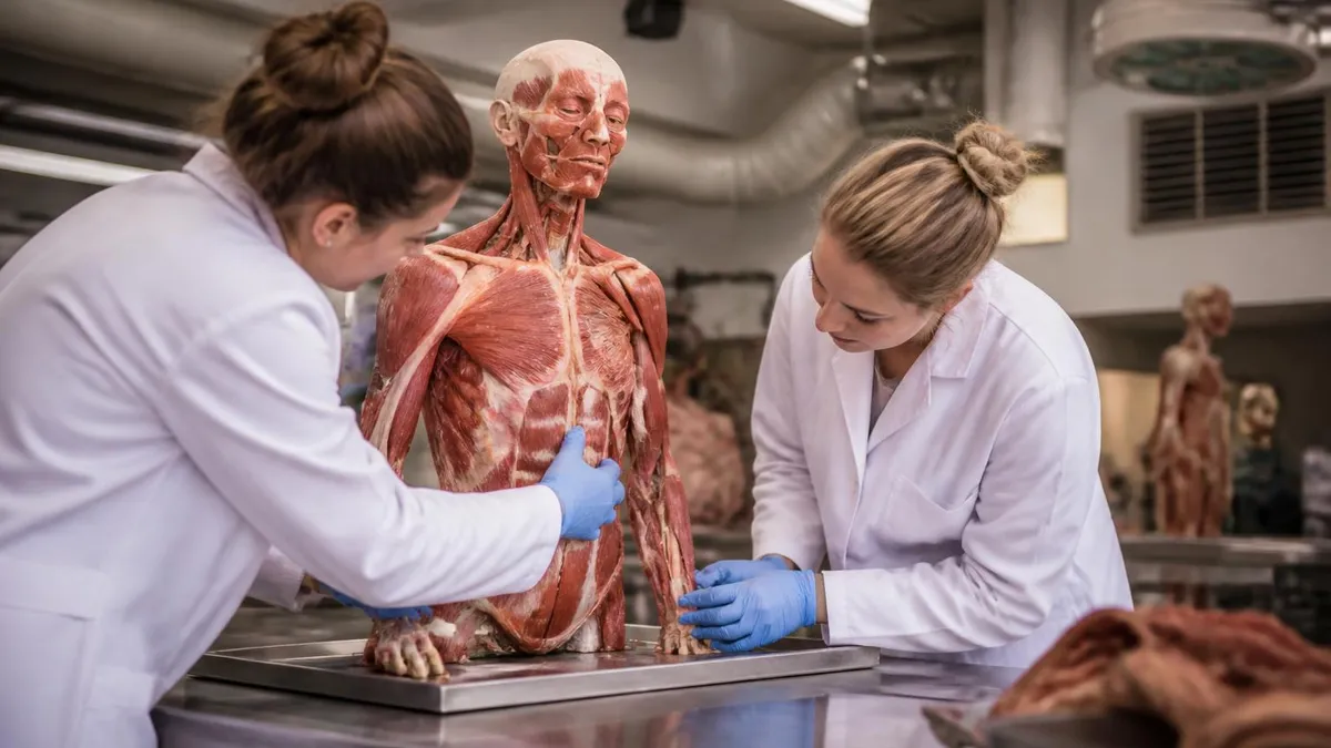
Human Anatomy and Physiology Book: The Complete Guide to Choosing, Using, and Mastering the Best Textbooks for 2026

Best Way to Learn Anatomy and Physiology: A Complete Student Guide
About the Author

Educational Psychologist & Academic Test Preparation Expert
Columbia University Teachers CollegeDr. Lisa Patel holds a Doctorate in Education from Columbia University Teachers College and has spent 17 years researching standardized test design and academic assessment. She has developed preparation programs for SAT, ACT, GRE, LSAT, UCAT, and numerous professional licensing exams, helping students of all backgrounds achieve their target scores.