Pacemaker and MRI: A Complete Safety and Imaging Guide
Pacemaker and MRI safety guide covering MR-conditional devices, scanning protocols, screening, risks, and what patients and technologists need to know.
The relationship between a pacemaker and MRI was once considered an absolute contraindication, with cardiac implantable electronic devices (CIEDs) classified as a hard stop for magnetic resonance imaging. Over the past two decades, however, manufacturers, radiologists, and electrophysiologists have engineered a new generation of MR-conditional pacemakers and refined scanning protocols so that patients with cardiac devices can safely undergo MRI examinations when clinically necessary. Understanding the modern pacemaker and MRI workflow is essential for technologists, radiologists, and patients alike.
Roughly one in four pacemaker patients will need an MRI within four years of implantation, according to data from the Heart Rhythm Society. That statistic alone explains why hospitals can no longer treat cardiac devices as automatic disqualifiers. Instead, comprehensive screening, device interrogation, and programming workflows have replaced blanket refusals. The shift mirrors broader changes in how clinicians weigh diagnostic benefit against device-related risk, and it requires technologists to master a checklist far longer than the standard ferromagnetic screening process.
Modern MR-conditional pacemakers are designed with reduced ferromagnetic content, modified lead designs, and circuitry that tolerates the radiofrequency and gradient fields generated inside the bore. To learn more about the physics that makes these precautions necessary, review how does an MRI work for a foundational overview of the magnetic and RF principles involved. Without that physics background, the protective measures used during cardiac device imaging can feel arbitrary rather than physiologically driven.
The three primary hazards remain consistent across every guideline: heating of the lead tips that can damage myocardial tissue, induced currents that can trigger inappropriate pacing or arrhythmias, and mechanical force or torque on ferromagnetic components inside the generator. Each of these mechanisms scales with field strength, gradient slew rate, and specific absorption rate (SAR), which is why protocols often restrict scanning to 1.5T systems with normal-operating-mode SAR limits below two watts per kilogram.
Despite these constraints, the safety record for MR-conditional devices is strong. Multiple registry studies, including the MagnaSafe registry, demonstrate that adverse event rates remain below one percent when proper protocols are followed, even for some legacy non-conditional devices. That data has driven the FDA, the American Heart Association, and the American College of Radiology to publish consensus statements supporting MRI use in carefully screened pacemaker patients.
This guide walks through every dimension of the topic: device classifications, screening workflows, programming changes, monitoring requirements, post-scan checks, patient communication strategies, and registry-style exam content that technologists encounter on certification tests. Whether you are preparing for the ARRT MRI registry, training new staff, or counseling a patient before their first scan, the principles here form the foundation of safe, effective cardiac device imaging in 2026 and beyond.
By the end, you will understand not just the rules but the reasoning behind them, which is what separates a competent technologist from a reactive one. Safe pacemaker and MRI imaging depends on judgment as much as on checklists, and judgment requires understanding mechanism, evidence, and edge cases.
Pacemaker and MRI by the Numbers
Pacemaker Device Classifications for MRI
Devices tested and labeled by the manufacturer as safe under specific conditions, including field strength, SAR limits, scan duration, and anatomic coverage. Most modern pacemakers fall into this category and include both generator and lead system labeling.
Legacy devices or those with known ferromagnetic risks that should not enter the MRI environment. These include many pre-2008 generators and abandoned leads, which retain significant heating and torque risk regardless of programming changes.
Newer-generation devices approved for scanning at three Tesla under strict conditions. Approval requires updated lead configurations and confirmation that the entire system, not just the generator, meets 3T labeling requirements.
Older devices not labeled MR-conditional but scanned off-label in select institutions following the MagnaSafe protocol. Requires institutional review, written consent, and a higher level of monitoring during and after the exam.
Patients with a conditional generator paired with non-conditional leads, or vice versa. These hybrid systems do not qualify as conditional and must be evaluated as non-conditional with appropriate caution and case-by-case review.
The mechanisms that make pacemaker and MRI interactions dangerous fall into three physical categories: magnetic, electrical, and thermal. Each interacts differently with the device and the patient, and understanding each one helps technologists explain risks accurately and respond appropriately when something unexpected happens. The static magnetic field, the gradient fields, and the radiofrequency pulses each create distinct hazards that scale differently with field strength, sequence design, and patient anatomy.
The static magnetic field, typically 1.5 or 3 Tesla, exerts force and torque on any ferromagnetic component. Modern pacemaker generators have been engineered to minimize ferromagnetic content, but reed switches, battery housing, and certain capacitors still respond to strong fields. The result can be unintended activation of asynchronous pacing modes, displacement of the device pocket, or mechanical stress on suture lines if the generator was implanted within the last six weeks. This is why most protocols require a healing window before scanning.
Gradient fields, the rapidly switching magnetic fields that encode spatial information, induce voltages in conductive leads. These induced currents can mimic intrinsic cardiac activity and confuse the device's sensing circuitry, potentially leading to inappropriate inhibition of pacing in pacemaker-dependent patients. Programming the device to an asynchronous mode such as DOO or VOO before the scan eliminates this risk by ensuring the device paces at a fixed rate regardless of what it senses.
Radiofrequency energy is the most discussed hazard because it produces heating at the lead tip. The tip is where the electrode contacts myocardial tissue, and even modest temperature rises can cause edema, fibrosis, or threshold changes that affect long-term pacing performance. For more context on how RF energy is used to generate signal, see what's the difference between MRI and CT scan, which contrasts MRI's RF-based imaging with the ionizing radiation used in computed tomography.
Specific absorption rate, expressed in watts per kilogram, is the regulated metric that protocols use to control RF exposure. Normal operating mode caps whole-body SAR at two watts per kilogram, and most MR-conditional pacemaker labeling requires this limit. First-level controlled mode allows up to four watts per kilogram but is generally avoided for cardiac device patients because the additional thermal margin is not worth the small diagnostic gain in most cases.
Lead length, geometry, and routing also influence heating. A lead that runs parallel to the long axis of the bore acts like an antenna at certain RF frequencies, concentrating energy at the tip. This is why some conditional labeling restricts scans of the thorax or upper extremities more tightly than lower extremity or brain scans. Technologists who understand this antenna effect can anticipate which exams require the most conservative parameter choices.
Finally, the device itself can be affected by cumulative exposure. Repeated scans can shorten battery life, change pacing thresholds, or alter sensing characteristics. These changes are usually subtle and reversible, but they reinforce the importance of post-scan device interrogation to document any parameter drift and adjust programming if needed.
Pacemaker and MRI Screening and Workflow
Screening begins long before the patient enters the MRI suite. The technologist verifies the device model, manufacturer, and implant date using the patient's device identification card, the medical record, or a manufacturer database query. Every component, including the generator and all leads, must be confirmed MR-conditional and matched to the labeled conditions for field strength and SAR.
Additional questions cover implant date, presence of abandoned or fractured leads, and any known device alerts or recalls. If documentation is incomplete, the scan is deferred until the electrophysiology team can interrogate the device and confirm its labeling. Skipping this step is the single most common cause of adverse events in pacemaker and MRI workflows.
Should a Pacemaker Patient Have an MRI?
- +Provides superior soft tissue contrast unavailable with CT for many neurologic and musculoskeletal questions
- +Avoids ionizing radiation, which is especially important for younger or frequently imaged patients
- +MR-conditional devices have a strong safety record with sub-one-percent adverse event rates
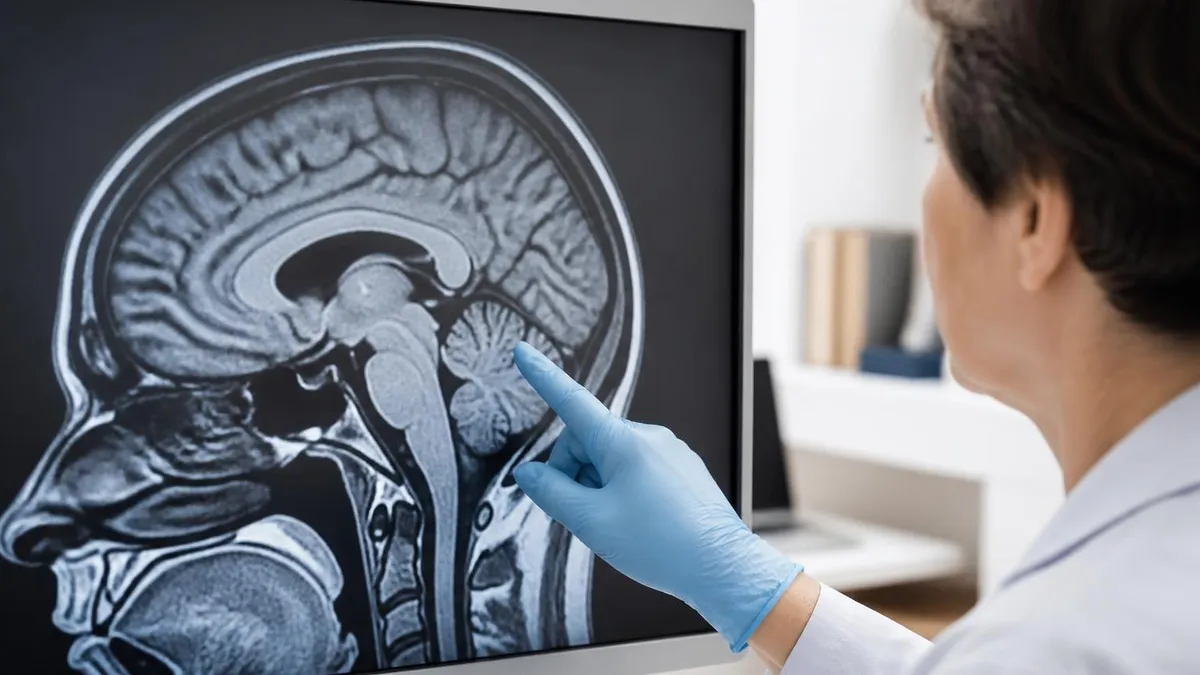
- +Enables diagnosis of stroke, multiple sclerosis, and tumors that influence cardiac and overall care
- +Modern protocols and registries support scanning even some non-conditional devices safely
- +Avoids repeat imaging or invasive alternatives when MRI is the appropriate modality
- −Requires extensive screening, programming, and electrophysiology coordination before each scan
- −SAR and sequence restrictions can limit image quality compared with unrestricted protocols
- −Risk of lead-tip heating, induced arrhythmias, or device reset remains nonzero
- −Post-scan device interrogation adds time and cost to every examination
- −Pacemaker-dependent patients require continuous monitoring and trained clinical staff
- −Some legacy or mixed systems still cannot be scanned safely under any protocol
Pacemaker and MRI Pre-Scan Checklist
- ✓Verify device manufacturer, model, and serial number against the patient's device card
- ✓Confirm MR-conditional labeling for both generator and all implanted leads
- ✓Document implant date and ensure healing window of at least six weeks has elapsed
- ✓Check for abandoned, fractured, or epicardial leads that may disqualify the patient
- ✓Coordinate with electrophysiology for pre-scan device interrogation and reprogramming
- ✓Disable tachyarrhythmia therapies on combined pacemaker-defibrillator devices
- ✓Program asynchronous pacing for pacemaker-dependent patients during the scan
- ✓Verify scanner is set to normal operating mode with whole-body SAR under two watts per kilogram
- ✓Confirm continuous ECG, pulse oximetry, and visual monitoring are in place
- ✓Ensure ACLS-trained clinician and emergency equipment are immediately available
- ✓Restore baseline programming and perform post-scan device interrogation
- ✓Document any parameter changes, symptoms, or adverse events in the patient record
Conditional Does Not Mean Unconditional
An MR-conditional label only applies when every condition in the manufacturer's instructions for use is met. Field strength, SAR mode, scan duration, anatomic coverage, and lead configuration must all match the labeling. A single deviation, such as scanning the thorax when only the head is approved, voids the conditional status and exposes both patient and institution to risk.
Programming and monitoring form the operational backbone of any safe pacemaker and MRI workflow. Even with a fully conditional device, the scan should not begin until programming has been adjusted to mitigate the predictable risks of gradient-induced sensing errors and inappropriate therapy delivery. This is a collaborative process between MRI staff, electrophysiology, and sometimes anesthesia, and it requires clear documentation at every step.
For pacemaker-dependent patients, asynchronous modes such as DOO, VOO, or AOO are the standard. These modes deliver pacing at a fixed rate without regard to intrinsic activity, which prevents gradient noise from inhibiting necessary pacing. The rate is typically set above the patient's intrinsic rhythm to ensure capture, often around 80 beats per minute, though individual physiology and rhythm dictate the exact setting.
Non-dependent patients can often be programmed to ODO or OVO modes that disable pacing entirely during the scan. This eliminates the risk of inappropriate pacing while accepting that the patient's intrinsic rhythm will sustain them through the exam. The choice depends on documented intrinsic rate, prior Holter data, and the electrophysiologist's clinical judgment about how the patient will tolerate brief periods without backup pacing.
Defibrillator components, when present, must have all tachyarrhythmia detection and therapy turned off. Gradient noise is the primary culprit here because the rapid switching can mimic ventricular fibrillation on the device's sensing channel, leading to shock delivery during the scan. Disabling detection prevents this without removing the ability to restore therapies immediately after the patient exits the magnet.
Monitoring during the scan extends beyond simple visual observation. Three-lead ECG telemetry compatible with the magnet, pulse oximetry, and direct line of sight through the control room window are minimum standards. Some institutions add capnography for sedated patients. The goal is to detect any rhythm change, oxygen desaturation, or symptomatic event within seconds so that the sequence can be paused and the patient evaluated.
Personnel requirements are equally explicit. An ACLS-trained clinician, often the electrophysiology nurse who performed the pre-scan programming, must remain in the control area for the duration of the scan. A physician familiar with the device must be reachable within minutes. Emergency equipment including an external pacing and defibrillation system stays outside the magnet room because none of it is MR-safe.
After the scan, the device is interrogated again to verify pacing thresholds, lead impedance, and battery voltage. Parameter drift is documented and compared with pre-scan values. Significant changes prompt closer follow-up, and minor changes are logged for trending across future scans. This closes the loop and ensures that pacemaker and MRI imaging remains safe not only during the exam but across the device's full service life.
Patients with abandoned, fractured, or capped leads that remain in the body are generally excluded from MRI, even if the active device is MR-conditional. Abandoned leads can still heat at the tip and have not been tested under conditional labeling. Confirm lead status with chest radiography and device records before scheduling, and consult electrophysiology if any uncertainty exists.
Patient communication is often the most underrated piece of pacemaker and MRI workflows, yet it determines whether the patient arrives prepared, calm, and able to comply with breath-hold and stillness requirements. Most pacemaker patients have been told for years that MRI is forbidden, and that message persists even after they receive an MR-conditional device. Reorienting them requires patience, accurate information, and written materials they can review at home.
The conversation begins at scheduling. Schedulers should confirm device type, gather the patient's device card information, and explain the additional steps such as pre-scan electrophysiology programming. Patients are told to bring their device identification card, a list of current medications, and any prior imaging on disc. They are also instructed to arrive earlier than a standard MRI appointment because programming and monitoring setup add 30 to 60 minutes.
On the day of the exam, the technologist reviews the screening form line by line, even for patients who have had prior MRIs. Devices can be upgraded, leads added, or recalls issued in the interval since the last scan, and a fresh review catches changes that paperwork alone might miss. To help patients understand why contrast may or may not be used in their study, share MRI with or without contrast: what to expect as a pre-visit resource.
Patients are reassured that the scan itself will feel like any other MRI, with the same loud knocking and the same enclosed bore. The difference, they are told, lies in the preparation and monitoring rather than the scan experience. This framing reduces anxiety and helps patients focus on staying still rather than worrying about their device.
Clear instructions about what to do if they feel something unusual are essential. Patients are taught to use the squeeze ball for any chest discomfort, palpitations, lightheadedness, or unusual warmth at the device site. They are reassured that minor sensations are common and not necessarily dangerous, but that the team prefers to evaluate rather than ignore symptoms. This empowers the patient without creating false alarm.
After the scan, patients are informed about post-scan device interrogation and any programming changes made. They are reminded to follow up with their electrophysiologist as scheduled and to report any new symptoms in the days following the exam. Written discharge instructions reinforce the verbal review and give caregivers something to reference at home.
Finally, technologists and nurses should be prepared to address common myths. Many patients believe that MRI will magnetize their device permanently, drain the battery in one sitting, or pull the generator out of the chest. Calm, accurate explanations rooted in mechanism rather than reassurance alone build trust and improve compliance not only for this scan but for any future imaging the patient may need.
Practical preparation for pacemaker and MRI scans benefits from a few habits that experienced technologists develop over time. First, build a relationship with your electrophysiology team. The smoother the communication, the faster the workflow, and the less likely small errors are to slip through. Joint case reviews, shared screening forms, and shared documentation templates pay dividends across hundreds of scans annually and reduce the cognitive load on both teams when complex cases arrive.
Second, maintain a current list of approved devices and conditional labeling at your scanner console. Manufacturers update labeling regularly, and a device that was non-conditional last year may have received an updated approval through firmware changes. The MRISafety.com database, manufacturer portals, and ACR resources are reliable starting points, and many departments keep printed quick-reference sheets at the screening desk for fast lookup.
Third, rehearse emergency procedures with the full team at least quarterly. The actions required if a patient becomes symptomatic in the bore must be automatic. Quench-button responsibilities, code blue procedures adapted for the MRI environment, and rapid removal techniques should all be drilled. Cardiac device patients raise the stakes because pacing failure or arrhythmia can deteriorate rapidly, and there is no time to read a manual when seconds matter.
Fourth, document everything. Pre-scan interrogation values, programming changes, monitoring observations, scan parameters, and post-scan interrogation values all belong in the permanent record. This protects the patient, supports continuity of care, and provides a defensible record if questions arise later. Many institutions use a structured CIED MRI checklist that becomes part of the imaging report.
Fifth, recognize when to say no. Not every pacemaker patient is a good MRI candidate, even with conditional labeling. Abandoned leads, recent implants within the healing window, unstable rhythms, or unavailable device interrogation are legitimate reasons to defer the exam and pursue alternative imaging. Saying no is sometimes the safest professional decision, and confident decision-making protects both the patient and the team's credibility.
Sixth, stay current with guidelines. The Heart Rhythm Society, ACR, and ASE publish updates, and registry data continues to refine which non-conditional devices can be safely scanned under defined protocols. Continuing education, journal clubs, and manufacturer webinars are inexpensive ways to keep your knowledge sharp. Registry exams reflect these updates, so test preparation also serves as ongoing professional development for the technologist.
Finally, treat each pacemaker patient as an individual rather than a category. Two patients with the same device model may have very different rhythms, anxieties, and clinical contexts. The protocol provides the framework, but the technologist's judgment, communication, and attention to detail determine whether the scan goes smoothly. That blend of standardization and individualization is what modern pacemaker and MRI imaging requires, and it is what registry exams ultimately try to measure.
MRI Questions and Answers
About the Author
Medical Laboratory Scientist & Clinical Certification Expert
Johns Hopkins UniversityDr. Sandra Kim holds a PhD in Clinical Laboratory Science from Johns Hopkins University and is certified as a Medical Technologist (MT) and Medical Laboratory Scientist (MLS) through ASCP. With 16 years of clinical laboratory experience spanning hematology, microbiology, and molecular diagnostics, she prepares candidates for ASCP board exams, MLT, MLS, and specialist certification tests.