
MRI Pituitary: Protocol, Sequences and Reporting Guide 2026 September
🗨️ MRI pituitary protocol covers thin slice T1, T2, dynamic post-contrast sequences, microadenoma detection and reporting tips for techs and trainees.
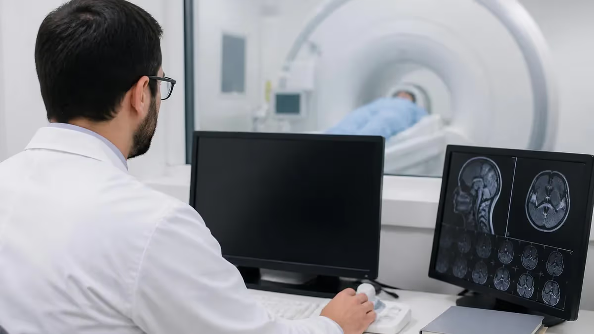
The pituitary gland sits in the sella turcica, a bony pocket no wider than a small coin. Imaging it well takes patience and a tight protocol. An MRI pituitary study is the first line for headache with visual loss, suspected Cushing disease, prolactin elevation, panhypopituitarism and acromegaly.
Cross-sectional resolution matters here. A 3 mm cut through the gland can hide a 4 mm microadenoma that a 2 mm cut would catch. That is why dedicated pituitary protocols differ from a routine brain MRI in sequence count, slice thickness and contrast timing.
Most centers run the sella study at 1.5 T or 3 T. The higher field gives sharper detail in the cavernous sinus and infundibulum, but it also brings susceptibility artifact from dental work and air in the sphenoid sinus. You learn to weigh those trade-offs at the console.
New techs often ask whether a brain scan is enough. It is not. A standard brain protocol uses 5 mm slices and skips dynamic enhancement. Tiny adenomas hide. The radiologist then has to ask for a repeat, the patient comes back, and the workflow takes a hit.
Pituitary MRI by the Numbers
Dynamic Phase is Critical
Dynamic post-contrast T1 with 15-second phases is the single most important sequence for microadenoma detection. Skip it and small lesions hide. Detection drops by half when dynamic imaging is omitted in series comparing standard versus dedicated pituitary protocols.
Why the pituitary needs its own protocol
The gland averages 10 mm by 8 mm in an adult. Microadenomas measure under 10 mm by definition, with most clinically relevant ones in the 3 to 7 mm range. Macroadenomas push above 10 mm and often distort the optic chiasm, the cavernous sinus walls or the sellar floor.
Resolution that reveals a 4 mm lesion needs thin slices, a small field of view and high signal to noise. Standard brain MRI cannot deliver that mix. Pituitary MRI uses 2 to 3 mm slices, a 16 to 18 cm field of view and a 256 or 320 matrix. The voxel is small. The contrast curve matters.
Dynamic post-contrast imaging is the second pillar. The normal gland enhances early because of its rich portal blood supply. Adenomas enhance later. By scanning the gland every 15 to 30 seconds after contrast injection, you catch the window where the adenoma still looks dark while the normal tissue is already bright.
That contrast difference is sometimes the only sign of a small lesion. Skip the dynamic, and the study can read as normal even when a microadenoma sits there. The detection rate drops by half in some series when the dynamic phase is omitted.
Standard sequence list
Most workflows include sagittal T1, coronal T1, coronal T2, coronal dynamic post-contrast T1 and delayed coronal and sagittal post-contrast T1. Slice thickness sits at 2 to 3 mm with no gap. Some centers add a 3D volumetric T1 SPGR or MP-RAGE for surgical planning.
Others add a coronal T2 SPACE or CISS for cisternal anatomy when arachnoid invasion is in play. The sequence list is short on paper but the timing window is tight in practice. A patient who moves during the dynamic loses the most diagnostic part of the study.
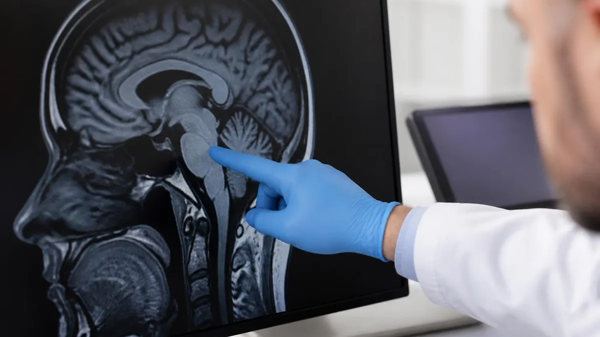
Pre-contrast T1 shows the posterior pituitary bright spot, a useful anatomic landmark. Loss of that bright spot is a clue in central diabetes insipidus. T2 helps differentiate cystic from solid and shows the optic chiasm well. Post-contrast highlights enhancement patterns and stalk position.
The stalk should be midline. A deviated stalk in a young patient with amenorrhea often points to a small adenoma on the opposite side. The reader needs to compare stalk position across coronal slices, not just one image.
Standard Sequence Stack
Anatomic overview that shows the posterior pituitary bright spot, the stalk in the mid-sagittal plane, and the sellar floor relationship with the sphenoid sinus. Slice thickness 2-3 mm, no gap, full coverage from anterior to posterior gland edges.
- Slice thickness: 2-3 mm
- Field of view: 16-18 cm
- TR/TE: 450-600 / 10-15 ms
Thin coronal slices perpendicular to the sellar floor, covering the entire gland from anterior to posterior. T2 distinguishes cystic from solid components and highlights the optic chiasm against CSF. Matrix 256 or 320 for high resolution.
- Slice thickness: 2-3 mm
- Matrix: 256 or 320
- Plane: Perpendicular to sellar floor
Six to eight phases at 15 to 30 second intervals captures the enhancement curve difference between normal gland (rapid uptake) and adenoma (delayed uptake). The single most important sequence for microadenoma detection. Injection rate 2 ml/sec.
- Phases: 6-8
- Interval: 15-30 sec
- Injection rate: 2 ml/sec
Coronal and sagittal T1 sequences after the dynamic, evaluating cavernous sinus invasion, suprasellar extension, optic chiasm contact and dural enhancement. Helpful for Knosp grading and surgical planning.
- Sequences: Coronal + sagittal T1
- Timing: 3-5 min post-injection
- Purpose: Knosp grading
Dynamic contrast: timing is everything
For the dynamic phase, the tech preloads the IV with 0.1 mmol per kg of gadolinium-based contrast, usually gadobutrol or gadoteridol. The injection runs at 2 ml per second. Scanning starts at the moment of injection or just before.
Most protocols capture six to eight phases at 15 second intervals. The normal gland lights up within 30 to 60 seconds. Microadenomas usually peak later. Some lesions, especially Rathke cleft cysts, never enhance.
One tip from the floor. Brief the patient before the scan. Tell them the contrast may feel cool. Tell them the scanner will be loud during the dynamic. Tell them to breathe gently and not to swallow if possible. The dynamic is short, maybe 90 seconds total, but a swallow during that window can ruin the loop.
Field Strength and Coil Comparison
Reliable workhorse for pituitary imaging. Less susceptibility artifact at the sphenoid sinus air-bone interface, which makes it the preferred field strength for transsphenoidal post-op follow-up. Slightly lower microadenoma detection compared with 3 T, but still sensitive for clinically relevant lesions above 4 mm. Most community hospitals run pituitary studies at this field strength. Coil: 32-channel head coil standard. Sequences identical to 3 T but with slightly thicker slices possible.
Field strength and coil choice
3 T offers higher signal and finer detail. Microadenoma detection can rise by 10 to 15 percent on 3 T compared with 1.5 T in some series. But 3 T also brings more susceptibility artifact at the sphenoid sinus interface.
The gland sits right above that air space. If the patient has had transsphenoidal surgery, the post-op artifact may be worse at 3 T. Many neuro radiologists prefer 1.5 T for post-op follow-up and 3 T for the initial workup. There is no single right answer.
Coil choice also matters. A standard 32-channel head coil works well. Some centers use a smaller surface coil for the pituitary alone, which boosts local signal but reduces coverage of the rest of the brain.
If you suspect a macroadenoma with suprasellar extension, you want the full head coil to image the chiasm, optic tracts and frontal lobes. Compromising coverage for signal is rarely the right trade.
Sudden severe headache with vision loss requires urgent imaging. T1 hyperintensity from methemoglobin and susceptibility weighted imaging help identify hemorrhage in an adenoma. The dynamic phase is less critical in this setting because the diagnosis hinges on the bleed, not subtle enhancement.
Common indications
The referral list is shorter than you would think. Hyperprolactinemia, Cushing disease, acromegaly, hypopituitarism, visual field defect, severe headache, suspected apoplexy and follow-up of known adenoma. Each indication shifts the protocol slightly.
For Cushing, the radiologist needs the highest sensitivity for small ACTH-secreting adenomas, so the dynamic is critical. For acromegaly, the lesion is usually larger but cavernous sinus invasion drives surgical planning.
For prolactinoma, response to medical therapy matters so a baseline study with stalk position and cavernous sinus involvement is key. See our pituitary microadenoma MRI page for sequence-specific tips.
Pituitary apoplexy is the emergency case. A patient comes in with sudden severe headache, vision loss and possibly altered consciousness. The radiologist looks for hemorrhage or infarction in an adenoma. T1 will show high signal if there is methemoglobin. Susceptibility weighted imaging adds value here.
Pituitary MRI Report Checklist
- ✓Gland size in three dimensions
- ✓Lesion size, location, signal characteristics
- ✓Stalk position (midline, deviated, thickened)
- ✓Cavernous sinus invasion with Knosp grade 0-4
- ✓Suprasellar extension and optic chiasm contact
- ✓Sellar floor integrity and clivus signal
- ✓Comparison with prior study, numeric change
- ✓Post-contrast enhancement pattern and timing
Reading the study: what the report needs
A clean report covers gland size, lesion size, lesion location within the gland, enhancement pattern, stalk position, cavernous sinus invasion, suprasellar extension, optic chiasm contact and sellar floor integrity.
Knosp grade is used for cavernous sinus invasion. Grade 0 means no invasion, grade 4 means the carotid is encased. Surgeons rely on Knosp grade to plan the approach. Hardy classification covers sellar floor and suprasellar extent.
Be specific about size. A 6 by 4 by 5 mm lesion in the left posterior gland is more useful than a vague mention of a small adenoma. The next reader needs that number for comparison on follow-up. Small adenomas can change slowly.
Pituitary MRI Strengths and Limits
- +Excellent soft tissue contrast for sellar anatomy
- +Dynamic contrast detects sub-centimeter adenomas
- +No ionizing radiation, safe for repeat follow-up
- +Multi-planar imaging shows chiasm and cavernous sinus
- +Surgical planning data from a single study
- −Susceptibility artifact from sphenoid sinus air
- −Long scan time (25-35 minutes) for anxious patients
- −Post-op packing material confounds early follow-up
- −Contrast contraindicated in severe renal impairment
- −Less sensitive for calcifications than CT
Pitfalls and artifacts
Several normal variants mimic adenoma. Pars intermedia cysts sit between the anterior and posterior lobes and look like small T2 bright spots with no enhancement. Cleft cysts can be large and may compress the gland.
They do not enhance internally, but they may have a thin enhancing rim. Pituitary hyperplasia in young women or postpartum can mimic enlargement. The gland is larger but enhances homogeneously and the stalk stays midline.
Susceptibility artifact from the sphenoid sinus air-bone interface is the most common technical pitfall. It distorts the inferior gland and can mimic or mask a lesion. Higher bandwidth helps. Lower echo time helps.
Fat saturation can sometimes fail near the sella because of field inhomogeneity, giving residual bright signal that confuses the dynamic. Spectral fat sat tends to fail more at 3 T near air spaces. SPAIR or STIR may work better in tough cases.
Pediatric pituitary MRI
Children come in for short stature, precocious puberty, central diabetes insipidus or visual symptoms. Protocols match the adult version but slice thickness drops to 1.5 to 2 mm because the gland is smaller. Sedation may be needed for younger children.
The posterior pituitary bright spot is more conspicuous in kids. Ectopic posterior pituitary is a classic pediatric finding tied to growth hormone deficiency, with the bright spot appearing at the median eminence rather than the sella floor.
Craniopharyngioma is the most common suprasellar mass in children. It shows a mix of cystic and solid components and often has calcifications, which MRI does not show well. CT pairs with MRI for craniopharyngioma workup.
Germinoma and Langerhans cell histiocytosis affect the stalk and can present with diabetes insipidus and a thickened pituitary stalk. The differential narrows quickly with clinical context.
Post-operative imaging
Transsphenoidal surgery leaves packing material in the sella that enhances variably and can mimic residual tumor in the early post-op period. Most centers wait three months for a baseline post-op MRI to let the packing change resolve.
After that, residual or recurrent disease shows as a true enhancing nodule with mass effect or progressive growth on follow-up. Compare with the immediate post-op image whenever possible.
If the patient had radiation therapy after surgery, expect gradual gland atrophy and possible empty sella on long-term follow-up. New hormonal deficits years after radiation are common and reflect slow hypothalamic and pituitary damage.
Tips for radiology techs and trainees
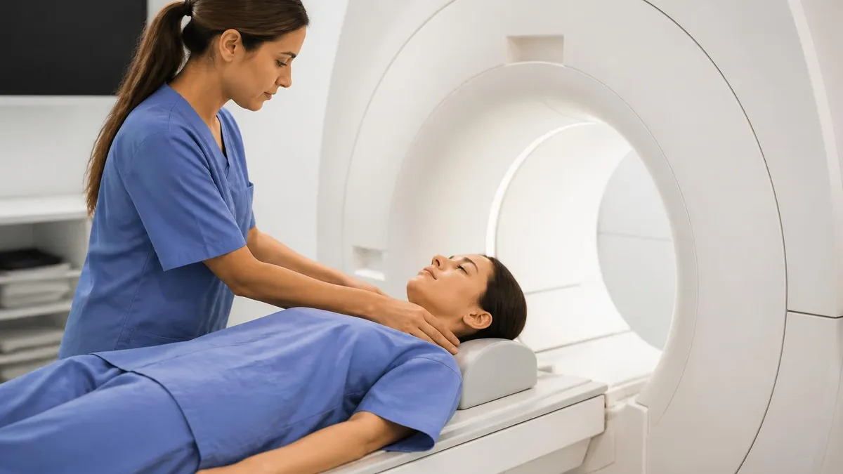
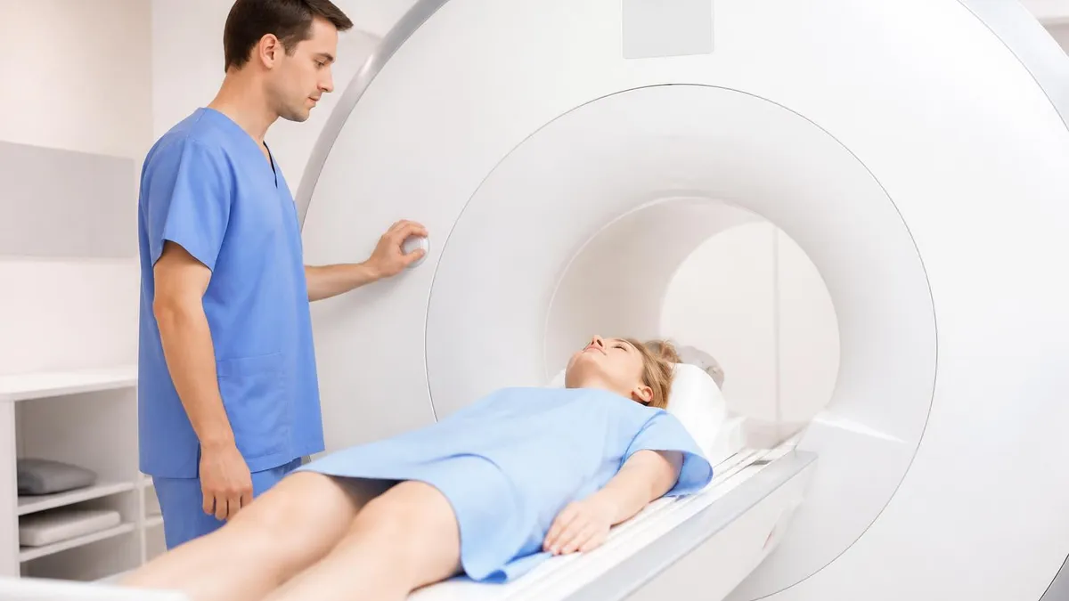
Position the patient comfortably. Use foam pads to reduce motion. Briefing matters. Patients with visual field defects often have anxiety and the magnet bore can feel claustrophobic. Pre-load the IV before positioning so the dynamic starts on time.
Set the saturation bands away from the sella. Check the localizer carefully. The coronal slices should be perpendicular to the sellar floor, not the AC-PC line. A coronal cut parallel to the brainstem misses the gland.
For trainees writing the report, follow a checklist. Gland size. Lesion. Stalk. Cavernous sinus. Chiasm. Sphenoid sinus. Clivus. Each item gets a line. Comparison study reviewed and changes noted in numbers.
Practice and certification
For MRI techs preparing for certification (ARRT MRI or equivalent), pituitary imaging questions cover sequence selection, slice thickness, contrast timing and safety. Knowing the difference between dynamic and delayed phase shows up often.
Recognizing posterior pituitary bright spot anatomy comes up too. Practice with question banks that include neuro and contrast pharmacology sections. Reviewing the MRI brain protocol guide alongside pituitary makes the comparison clearer.
Safety and contraindications
Standard MRI safety screening applies. Cardiac pacemakers, neurostimulators, cochlear implants and certain aneurysm clips remain contraindications unless documented MR-conditional.
The patient should fill out the screening form in advance, but tech verification at the door is still required. Metal in the eyes, especially in patients with a welding history, calls for a screening orbital CT before the scan. The magnet pulls.
Contrast safety is a separate concern. Gadolinium is generally well tolerated, but patients with severe renal impairment face a risk of nephrogenic systemic fibrosis with older agents. Macrocyclic agents like gadobutrol and gadoteridol have a far lower risk profile.
Allergic reactions to gadolinium are rare, roughly 1 in 10,000 for moderate to severe reactions. Pre-medication with steroids and antihistamines is used for patients with prior reactions.
Comparing MRI with other modalities
CT plays a role when MRI is contraindicated. CT shows calcifications well, which matter in craniopharyngioma. CT also shows the sellar floor bony anatomy that surgeons need for the transsphenoidal approach.
For functional imaging, gallium-68 DOTATATE PET-CT can find ectopic ACTH sources in Cushing disease when pituitary MRI is negative. Inferior petrosal sinus sampling is the gold standard for confirming a pituitary source of ACTH.
For visual field testing, formal perimetry pairs with MRI to grade chiasm involvement. A patient with bitemporal hemianopia and a suprasellar mass needs both the structural read and the field map.
Putting it together
A good pituitary MRI study starts before the patient hits the table. The referring physician should send the clinical question. That context lets the tech load the right protocol.
Default brain pituitary settings without the dynamic give a slower turnaround and lower yield. The radiologist reads the study in about 15 minutes when the protocol is clean and the patient cooperated.
Quality control on pituitary MRI is mostly about the dynamic loop. Check that the injection timing matched the sequence start. Check that the slice positioning covered the whole gland. Check that there is no obvious motion blur on the coronal series.
If any of those check boxes fail, the study needs a rerun. Pituitary MRI rewards careful technique. The lesions are small, the anatomy is tight, and the consequences of a miss reach the operating room.
Common questions techs hear
Patients often ask why the scan takes so long, why there is a separate set of pictures after the injection, and what the noise means. Honest, short answers help. The dynamic phase needs a tight sequence of images right after contrast hits the gland. The noise is normal. The clicks and hums are the gradients switching.
Another question: why no metal. Even small metal objects can heat up or move in the magnet. Hair clips, jewelry, underwire bras, glasses. All come off. Tattoos with metallic ink are usually fine but rarely cause local warmth. Permanent makeup is the same. A brief skin check before the scan picks up the rare reaction.
Patients with claustrophobia struggle with closed-bore systems. Open-bore or wide-bore magnets help but are not as sharp for pituitary work. A short course of oral anxiolytic from the referring physician can make the scan possible. Music in the headphones helps too. A patient who can lie still gets a better picture.
Workflow for the busy department
A high-volume neuroradiology service might run six to ten pituitary studies a day. Batching them together at certain times of the day helps the techs build muscle memory for the protocol. The dynamic phase becomes second nature. Mistakes drop. The radiologist reads faster.
Storing protocol templates in the scanner is essential. Manual entry of slice positioning and timing wastes time and introduces error. Most modern scanners have a pituitary protocol preset that the tech just loads and tweaks for the patient. Tweaks include adjusting the field of view for the patient's head size and the saturation bands for any orthodontic hardware.
Reporting templates speed up the radiologist. A pituitary report has the same backbone every time. Pre-filled headings for gland measurements, lesion description, stalk, cavernous sinus, suprasellar extension and comparison cuts read time in half. Voice recognition with smart templates is now the norm.
MRI Questions and Answers
About the Author

Medical Laboratory Scientist & Clinical Certification Expert
Johns Hopkins UniversityDr. Sandra Kim holds a PhD in Clinical Laboratory Science from Johns Hopkins University and is certified as a Medical Technologist (MT) and Medical Laboratory Scientist (MLS) through ASCP. With 16 years of clinical laboratory experience spanning hematology, microbiology, and molecular diagnostics, she prepares candidates for ASCP board exams, MLT, MLS, and specialist certification tests.
Join the Discussion
Connect with other students preparing for this exam. Share tips, ask questions, and get advice from people who have been there.
View discussion (8 replies)