What Is Contrast for MRI? A Plain-English Guide to Gadolinium 2026 July
What is contrast for MRI? A clear guide to gadolinium-based contrast agents — how they work, when used, safety, NSF risk, cost, pregnancy. ✍🏼

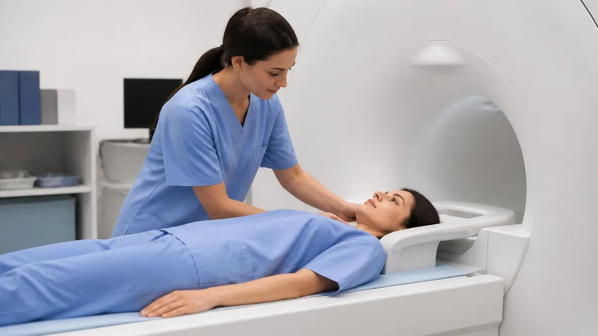
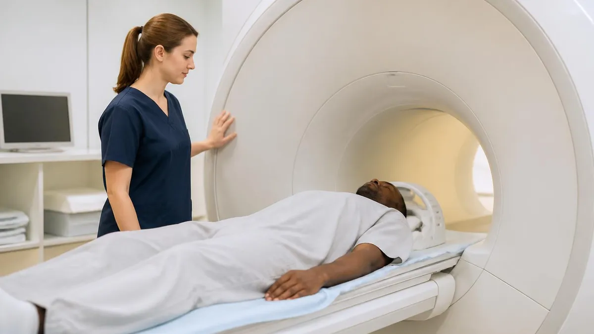
You scheduled an MRI and the order says "with and without contrast". The technologist starts an IV. A small syringe of clear liquid goes in. Suddenly your scan looks different on the screen — brighter spots, sharper edges, things the radiologist couldn't quite see before. That liquid is gadolinium, and it's the whole reason your doctor asked for contrast in the first place.
MRI contrast is not the same as the iodine dye used in CT scans. It works differently, it's processed by the body differently, and it carries a different (and much smaller) risk profile. Still, if you're being asked to get one, you probably want to know what's actually being injected, why it matters for your diagnosis, and whether you should worry about anything.
This guide walks through what gadolinium-based contrast agents (GBCAs) are, how they make MRI images more useful, when radiologists actually need them, and what the real safety picture looks like — including the rare-but-serious nephrogenic systemic fibrosis (NSF), the modern shift toward macrocyclic agents, and what to do if you're pregnant or breastfeeding. By the end you'll know exactly what the technologist is about to inject, and why.
MRI contrast is a gadolinium-based liquid injected into a vein in your arm. It makes certain tissues — tumors, inflamed areas, blood vessels, MS lesions — light up on T1-weighted images so the radiologist can see them clearly. It contains no iodine, and serious reactions are very rare (less than 0.1%). Most people feel nothing at all.
What MRI contrast actually is
The dye your technologist injects is called a gadolinium-based contrast agent — GBCA for short. Gadolinium is a rare-earth metal. In its raw form it's toxic, so the manufacturers chemically lock each gadolinium atom inside a larger molecule (the chelating cage) that the body can't break apart. The whole package is excreted intact, mostly through the kidneys, within about 24 hours.
You may hear brand names tossed around in the imaging center. The big ones in the U.S. are Dotarem (gadoterate), Gadavist (gadobutrol), ProHance (gadoteridol), MultiHance (gadobenate), Eovist (gadoxetate, for liver work), and Magnevist (gadopentetate — older, mostly retired now). The first three are macrocyclic, which is the safer modern class. We'll come back to that.
Worth saying up front: this is not the iodinated contrast used in CT scans. Iodine and gadolinium are completely different elements with completely different chemistry. An iodine allergy does not predict a gadolinium reaction, and vice versa. If the front desk asks about "contrast allergies" make sure they know which scan you had a problem with.
MRI contrast by the numbers
How gadolinium makes images brighter
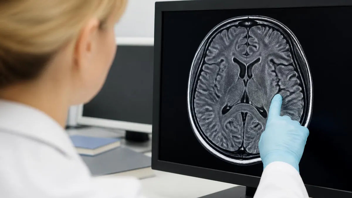
This is the part textbooks tend to overcomplicate. The short version: MRI doesn't image gadolinium directly. It images water — specifically the hydrogen protons inside water molecules. What gadolinium does is change how those protons behave around it.
Every tissue in your body has its own characteristic relaxation times — T1 and T2. T1 is how fast protons release energy back to the surrounding tissue after the scanner pulses them. Gadolinium is paramagnetic, meaning it has unpaired electrons that wobble nearby protons and dramatically shorten T1. Tissues that soak up contrast appear brighter on T1-weighted sequences.
Why do some tissues soak it up more than others? Three reasons: blood supply, vascular leakiness, and breakdown of the blood-brain barrier. A tumor has chaotic, leaky vessels — so gadolinium pours in. An MS lesion in an active flare disrupts the blood-brain barrier — so gadolinium leaks across. A bone bruise is just bruised, with normal vessels — so it barely enhances at all. The pattern of brightness is a fingerprint, and radiologists read those fingerprints for a living.
The technologist typically scans you first without contrast, injects, then runs the same sequences again. The radiologist compares the pre- and post-contrast images side by side. Anything that changed lit up because of gadolinium uptake. That's the diagnostic signal.
There's a timing element too. Some tissues enhance immediately and stay bright. Others enhance late, peaking 20 to 40 minutes after injection. Liver imaging in particular uses multiple time points — arterial phase, portal venous, delayed — because hepatocellular carcinoma and benign hemangiomas wash in and out on completely different schedules. Reading the timing curve is half the diagnosis.
What it is: Gadolinium-based contrast agent (GBCA), a paramagnetic metal in a chelated cage. What it does: Shortens T1 relaxation time so vascular and inflamed tissues glow on T1-weighted images. Who needs it: Patients being worked up for tumors, MS, infection, vascular disease, or anything where the radiologist needs to see active blood flow and tissue permeability. Who skips it: Most routine joint, spine, and back-pain scans — and anyone with severe kidney disease unless the benefit clearly outweighs the risk.
When contrast is needed (and when it isn't)
Contrast doesn't make every MRI better. For a lot of musculoskeletal work it adds nothing. The decision is driven by what the radiologist is actually looking for.
Brain MRI for suspected multiple sclerosis: almost always with contrast. Active demyelinating lesions break the blood-brain barrier and enhance brightly, which is how radiologists separate "old, quiet" plaques from "new, active" ones. The McDonald criteria for MS diagnosis depend on this distinction.
Brain MRI for headache or stroke workup: usually without. Stroke shows up beautifully on diffusion-weighted imaging, which doesn't need contrast. Most headache workups don't either.
Pituitary or sellar mass: with. Pituitary microadenomas are tiny and only visible because they enhance differently from the surrounding gland.
Spine MRI for radiculopathy or disc herniation: usually without. Discs and nerve roots show up clearly on standard T1 and T2 sequences. Add contrast if there's a history of prior back surgery (to separate scar tissue from recurrent disc), suspected infection, or a possible tumor.
Knee, shoulder, hip for sports injury: usually without. ACL tears, meniscal tears, rotator cuff pathology — all visible on standard sequences. The exception is MR arthrography where contrast is injected into the joint by a radiologist, which is a totally different procedure.
Liver, pancreas, kidney imaging for masses: with. Vascular enhancement patterns are the entire reason these studies are ordered. A liver lesion that washes in early and washes out late looks completely different from one that enhances slowly — and that pattern often tells you whether you're looking at cancer or a benign hemangioma.
Breast MRI for cancer screening or staging: with. Breast MRI without contrast is essentially useless. Cancers enhance rapidly and washout quickly; the timing curves are diagnostic.
A note on what you can ask. If your doctor ordered "MRI with and without contrast" and you're wondering whether the contrast portion is strictly necessary, that's a fair conversation to have before the scan — not after. It changes the cost, the scan time, and whether you need an IV. Most ordering physicians will explain the indication if you ask politely. If the answer is something like "we just always add contrast for this", push a bit further: many "routine" contrast orders survive on inertia rather than evidence.
When radiologists ask for contrast
Most primary and metastatic tumors enhance because of their abnormal, leaky vasculature.
- ▸Brain tumors — gliomas, meningiomas, metastases
- ▸Liver lesions — HCC, metastases, hemangiomas
- ▸Breast cancer staging and screening
- ▸Pituitary microadenomas
- ▸Soft-tissue sarcomas
Inflammatory tissue is hyperemic — extra blood flow means extra contrast uptake.
- ▸Active MS lesions in the brain and spine
- ▸Discitis and vertebral osteomyelitis
- ▸Brain abscess and meningitis
- ▸Inflammatory bowel disease (IBD)
- ▸Septic arthritis evaluation
Gadolinium-enhanced MR angiography shows arteries and veins without iodinated dye or radiation.
- ▸Renal artery stenosis screening
- ▸Aortic aneurysm follow-up
- ▸Carotid artery disease
- ▸Peripheral vascular disease
- ▸Hepatic vein and portal vein assessment
Contrast separates scar tissue (which enhances late or not at all) from active disease.
- ▸Post-discectomy back pain — scar vs recurrent herniation
- ▸Glioma recurrence vs treatment effect
- ▸Post-mastectomy surveillance
- ▸Bowel anastomosis evaluation
- ▸Cardiac surgery follow-up
How contrast is given during your scan
The whole administration is over in under a minute. Before you go into the scanner the technologist places an IV — usually in the antecubital vein at the inside of your elbow, sometimes in the back of the hand. They'll do the first set of "pre-contrast" sequences with the IV already in.
Partway through the scan, often without even moving you out of the bore, the technologist injects the gadolinium. Adult doses are weight-based: roughly 0.1 mmol per kilogram, which for most people works out to 10-20 mL of liquid. Some sequences (especially dynamic liver imaging) use a power injector that pushes the dose in at a controlled rate; others are pushed by hand over about 30 seconds, followed by a saline flush to clear the IV line.
You might feel a brief coolness traveling up your arm. A small number of people get a metallic taste or mild flushing. Both pass within a minute. After that the scanner runs the post-contrast sequences and you're done.
The IV comes out before you leave. There's nothing you need to do afterward except drink water — staying well hydrated helps your kidneys flush the gadolinium out, which is essentially complete in 24 hours for anyone with normal renal function.
Macrocyclic vs linear gadolinium agents
Macrocyclic GBCAs hold gadolinium inside a closed, cage-like ring. The cage is thermodynamically and kinetically stable, meaning the gadolinium ion almost never escapes before excretion. These are the modern standard.
Examples: Dotarem (gadoterate), Gadavist (gadobutrol), ProHance (gadoteridol).
Why they matter: Brain retention studies showed essentially no measurable gadolinium deposition in tissue. NSF risk is also lower. If you're getting repeated MRIs (multiple sclerosis surveillance, cancer follow-up, anything that means many contrast-enhanced scans over years) and you have a say in which agent is used — this is the class you want.
The safety picture — what's actually risky
For the vast majority of patients with normal kidney function, gadolinium contrast is one of the safer medical interventions in modern medicine. Acute reactions of any kind hit about 1-2% of administrations, and serious reactions (anaphylaxis-grade) come in well under 0.1%. Most reactions are mild — flushing, nausea, brief itching at the injection site.
The bigger conversation is about a rare condition called nephrogenic systemic fibrosis (NSF). NSF was first identified in 1997 and connected to gadolinium in 2006. It's a progressive scarring of the skin, joints, eyes, and internal organs. There is no reliable treatment. It only occurs in patients with severely impaired kidney function — almost exclusively those with eGFR below 30, with the highest risk in dialysis patients.
Two things changed the risk landscape after 2008. First, every U.S. imaging center now screens kidney function before giving GBCA to anyone with risk factors (age over 60, diabetes, hypertension, known kidney disease, transplant history). A serum creatinine and calculated eGFR are checked, and contrast is held if the number is too low.
Second, the field shifted toward macrocyclic agents, which carry roughly 1/100th the NSF risk of the older linear ones. The result: NSF has become vanishingly rare. The American College of Radiology now classifies modern macrocyclic agents as having essentially zero risk in patients with stable renal function above eGFR 30.
Gadolinium retention in the brain — first reported in 2014 — is a separate concern. Studies showed that small amounts of gadolinium can deposit in the dentate nucleus and globus pallidus after multiple contrast-enhanced MRIs, even in patients with normal kidneys. No clear clinical disease has been linked to it, but the finding drove the move to macrocyclic agents and tighter dosing protocols.
Special populations — what changes the conversation
Patients with kidney disease
If your eGFR is below 30, your radiologist will weigh the diagnostic need very carefully before giving contrast. In most cases they'll either use a smaller dose of a modern macrocyclic agent, switch to a non-contrast MR sequence (MRA without contrast is possible using time-of-flight techniques), or pick a different modality entirely. Dialysis patients can receive gadolinium if absolutely necessary, with dialysis scheduled within 24 hours to clear the agent — but it's avoided when possible.
Pregnancy
Gadolinium crosses the placenta. Animal data suggest possible effects on the developing fetus, and a large 2016 Canadian study found a small increase in inflammatory conditions and stillbirth among women who got gadolinium during pregnancy. Bottom line: contrast-enhanced MRI is avoided in pregnancy unless the diagnostic information is essential and can't be obtained any other way. Most pregnant patients get non-contrast MRI instead, which is itself considered safe.
Breastfeeding
You don't need to pump and dump. Less than 0.04% of an IV dose passes into breast milk, and of that, less than 1% is absorbed by the infant's gut. The American College of Radiology states that breastfeeding can continue uninterrupted after gadolinium. If you'd feel more comfortable waiting 24 hours, that's reasonable, but it isn't medically required.
Pediatric patients
Children get the same dose-per-kilogram and benefit from the same safety profile. Macrocyclic agents are strongly preferred. The biggest practical issue for kids is the IV placement and holding still, not the contrast itself — many pediatric MRIs use sedation.
Prior reactions
If you've had a previous reaction to a GBCA, the imaging center will pre-medicate you with steroids and antihistamines starting 12-13 hours before the scan. Switching to a different gadolinium agent class also reduces recurrence risk. People who reacted to iodinated CT contrast are not at higher risk for gadolinium reactions — different drug, different chemistry.
Tell your technologist before contrast
- ✓Any prior reaction to MRI contrast (gadolinium) — name the agent if you remember
- ✓Known kidney disease, dialysis, or a recent creatinine result
- ✓Pregnancy or possibility of pregnancy
- ✓Asthma or significant allergy history
- ✓Sickle cell disease or hemolytic anemia
- ✓Single kidney, kidney transplant, or recent kidney surgery
- ✓Medications, especially diuretics or anything affecting kidney function
- ✓Whether you've had multiple contrast-enhanced MRIs in the past year
Cost, insurance, and what to expect on the bill
Adding contrast to an MRI bumps the price by a meaningful amount. The exact number depends on the imaging facility, the contrast agent used, and how much of it the protocol calls for. Hospital outpatient imaging tends to be the most expensive; freestanding imaging centers are typically 30-60% cheaper for the same scan.
Typical added cost in the U.S. ranges from $100 to $500. Insurance usually covers contrast when there's a clinical indication documented on the order. Where people get caught is in two places: first, the contrast itself may be billed as a separate line item with its own copay; second, scans labeled "with and without contrast" use roughly twice the scanner time and may be billed at a higher CPT code than a standard MRI.
A few practical notes. Ask up front whether the imaging center is in-network for both the facility fee and the radiologist's professional fee — these are billed separately and the radiologist may be out-of-network even when the building isn't. If you're paying cash, call ahead and ask for the cash price; it's often dramatically lower than the billed rate. And keep the CPT code from your order — knowing whether you're scheduled for, say, an MRI brain without contrast (70551), with contrast (70552), or with and without (70553) makes it possible to compare prices and benefits accurately.
MRI contrast — the trade-offs
- +Dramatically improves detection of tumors, inflammation, and active disease
- +Required for accurate MS lesion staging and follow-up
- +Enables MR angiography without ionizing radiation
- +Distinguishes scar tissue from recurrent disease after surgery
- +Modern macrocyclic agents are extremely safe in normal kidney function
- +No iodine — safe for patients with iodine/CT contrast allergies
- +Quick clearance — out of the body in about 24 hours
- +No need to interrupt breastfeeding
- −Adds $100-$500 to the scan cost
- −Adds 15-20 minutes to scan time
- −Requires IV placement
- −Small risk of nephrogenic systemic fibrosis in advanced kidney disease
- −Gadolinium brain retention with repeated use (clinical significance unclear)
- −Avoided in pregnancy unless essential
- −Rare acute reactions (<0.1% serious)
- −Not all scans need it — unnecessary contrast is unnecessary cost
The bottom line on MRI contrast
If you've been told you need a contrast-enhanced MRI, the most useful thing to know is why. Ask your ordering doctor: what specifically are you trying to see that needs contrast? In most cases the answer is concrete — "I want to look for active MS lesions", "I want to characterize this liver mass", "I want to rule out a tumor". That answer also tells you whether the contrast is genuinely needed or whether it was added by default.
For the scan itself, expect a small IV, a brief cool sensation when the gadolinium goes in, and roughly twice the scan time of a non-contrast study. If you have normal kidney function and no history of contrast reactions, the procedure carries very little real risk. The radiology team is screening for everything that matters — kidney function, allergies, pregnancy — before they ever touch the syringe.
If you want to keep building on what you've learned about MRI machine basics or read more about MRI safety in general, those companion guides go deeper on the hardware and the non-contrast safety questions. And if you're studying for an MRI tech credential, the contrast pharmacology in this article is fair game on the registry — try a practice round to see how it sticks.
MRI Questions and Answers
About the Author
Medical Laboratory Scientist & Clinical Certification Expert
Johns Hopkins UniversityDr. Sandra Kim holds a PhD in Clinical Laboratory Science from Johns Hopkins University and is certified as a Medical Technologist (MT) and Medical Laboratory Scientist (MLS) through ASCP. With 16 years of clinical laboratory experience spanning hematology, microbiology, and molecular diagnostics, she prepares candidates for ASCP board exams, MLT, MLS, and specialist certification tests.
Join the Discussion
Connect with other students preparing for this exam. Share tips, ask questions, and get advice from people who have been there.
View discussion (6 replies)