How to Pass Time in an MRI: Complete Guide to Preparing for Your MRI Scan
Learn how to pass time in MRI scans with proven mental tricks, breathing techniques, and prep tips that make the experience faster and stress-free. 🗨️

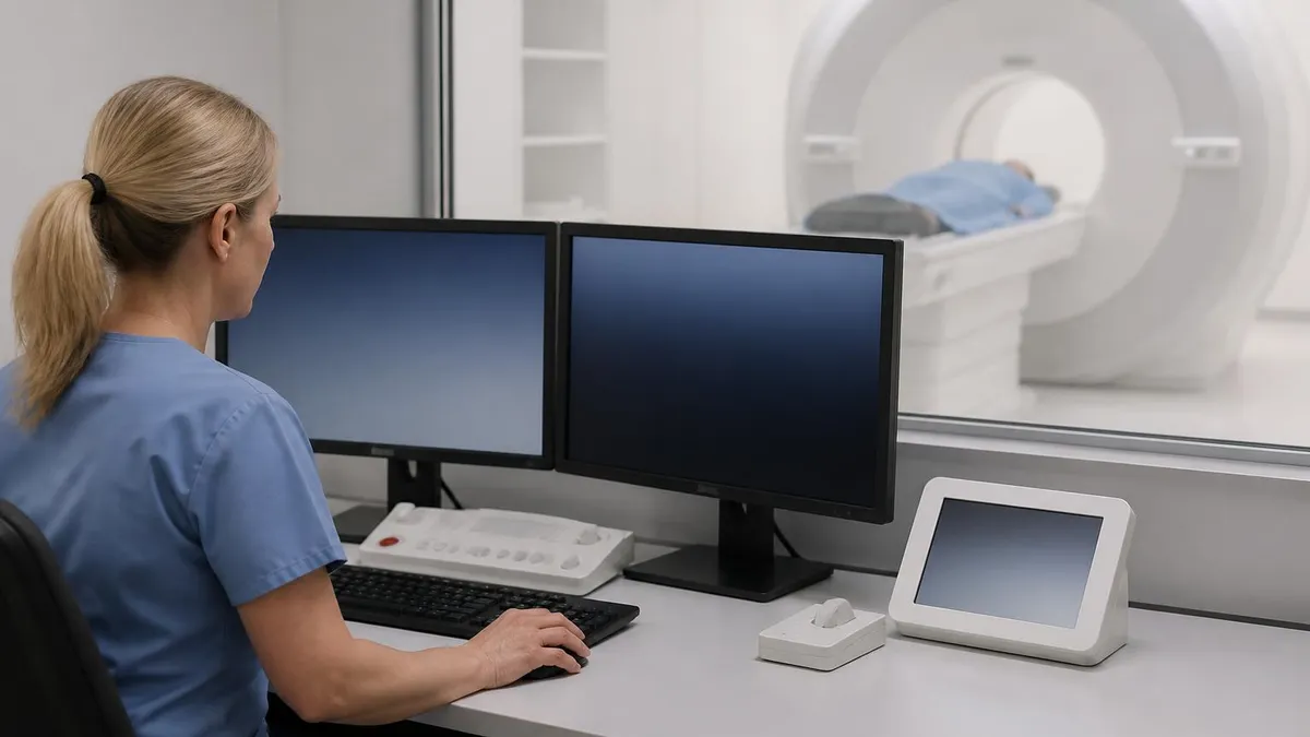
Figuring out how to pass time in mri appointments is one of the most common concerns patients raise before their scan, and for good reason. MRI exams can last anywhere from 15 minutes to over an hour, and the combination of confined space, loud noises, and the requirement to stay perfectly still creates a sensory experience unlike any other medical test. Understanding what to expect and preparing both mentally and physically transforms the experience from intimidating to manageable, even routine.
The good news is that the vast majority of patients complete their MRI without any significant distress. Modern scanners are faster, quieter, and more comfortable than older models, and radiology departments have refined their patient comfort protocols over decades of practice. Most facilities now offer headphones with music, mirrors that allow you to see outside the bore, and panic buttons that put you in immediate contact with the technologist throughout the entire scan.
Preparation begins days before your appointment, not minutes before you slide into the magnet. What you wear, what you eat, what medications you take, and how you mentally rehearse the experience all contribute to a smoother scan. Patients who arrive informed and relaxed produce clearer images, finish faster, and report dramatically better experiences than those who walk in cold without any preparation strategy in place.
This guide walks you through every aspect of MRI preparation, from the practical checklist of what to remove and bring with you to the psychological techniques that help time fly during the scan itself. You will learn how to handle contrast injections, what claustrophobia management options exist, and how to communicate with your technologist before and during the exam to make the entire process work in your favor.
We will also cover specific timing strategies that experienced MRI patients swear by, including mental exercises, visualization techniques, music selection tips, and breathing patterns that align with the scanner's rhythm. These methods are backed by patient feedback collected across thousands of scans and have helped countless people transform what could be an anxious hour into something they barely notice.
Whether this is your first MRI or your fifteenth, the strategies in this article will help you walk in confident, lie still without effort, and walk out wondering what you were worried about. By the time you finish reading, you will have a complete roadmap covering preparation, execution, and recovery, plus answers to the questions patients most frequently ask their radiologists and technologists about the experience.
MRI technology continues to evolve rapidly, with wide-bore systems, open MRI options, and faster sequences reducing scan times and patient discomfort year over year. Knowing what is available at your facility and asking the right questions before your appointment can dramatically change the experience. Let's start with the numbers behind MRI scans, then move through preparation, mental strategies, and the practical tips that experienced patients use to make their time in the bore pass quickly.
MRI Scans by the Numbers

What Happens During Your MRI Appointment
Check-In & Screening
Changing & Locker
Positioning
The Scan
Contrast Injection
Wrap-Up & Discharge
Preparing for an MRI starts with understanding what your specific scan requires. Different exams have different prep instructions, and the best place to start is the appointment confirmation paperwork your radiology department sent you. Abdominal MRIs often require fasting for four to six hours, MRCPs of the pancreas may require longer fasting, and pelvic MRIs sometimes require a partially full bladder. Cardiac and brain MRIs typically have no dietary restrictions at all, so always read your instructions twice.
Wardrobe choice matters more than most patients realize. Wear comfortable clothing without any metal components, including no zippers, no underwire bras, no metallic threads, and no embellishments. Athletic wear with metal eyelets, jeans with rivets, and many bras will need to come off. Most facilities provide gowns or scrubs, but arriving in metal-free clothing saves time and lets you keep your own clothes on, which many patients find more comfortable.
Medications usually continue as normal, but always confirm with your ordering physician. Diabetics on metformin may need to pause the drug for 48 hours after a contrast MRI if kidney function is borderline. Patients on anti-anxiety medications often take their prescribed dose 30-60 minutes before the scan, and your doctor can prescribe a one-time dose of lorazepam or diazepam specifically for the appointment if you have a history of claustrophobia or panic attacks.
Hydration plays a surprisingly important role, especially if contrast will be used. Drink water normally in the hours leading up to your scan unless instructed to fast. Well-hydrated patients have easier IV placements, better contrast clearance through the kidneys afterward, and generally feel more comfortable on the table. Avoid excessive caffeine on the day of the scan since it can amplify any underlying anxiety and make staying still more difficult than it needs to be.
The metal screening checklist is non-negotiable and exists to keep you safe. Pacemakers, cochlear implants, certain aneurysm clips, metallic foreign bodies in the eye, and some older joint replacements may make an MRI unsafe or require special protocols. Bring documentation of any implant you have, including the manufacturer card if available. Modern implants are usually MRI-conditional, meaning they are safe under specific scanner settings, but the radiologist needs the exact specifications in advance.
If you wear makeup, leave the heavy eye products at home. Some mascaras, eyeliners, and tattoo inks contain trace iron oxide that can heat up or distort images during the scan. Wash your face the morning of the appointment, skip the eye makeup, and remove nail polish if you are getting any kind of vascular imaging since pulse oximeters may be used. Hair products with glitter or metallic flakes should also be skipped on scan day.
For deeper context on how contrast affects your scan and what to expect when gadolinium is involved, the differences between contrast and non-contrast exams matter for your prep. Many patients also benefit from understanding the broader imaging landscape and how MRI compares to other tests their doctor might order, especially if they are choosing between scans.
MRI Practice Test Questions
Prepare for the MRI - Magnetic Resonance Imaging exam with our free practice test modules. Each quiz covers key topics to help you pass on your first try.
MRI Knowledge
MRI Exam Questions covering Knowledge. Master MRI Test concepts for certification prep.
MRI Physics
Free MRI Practice Test featuring Physics. Improve your MRI Exam score with mock test prep.
MRI Anatomy and Pathology
MRI Test Prep for MRI Anatomy and Pathology. Practice MRI Quiz questions and boost your score.
MRI Anatomy and Positioning
MRI Questions and Answers on MRI Anatomy and Positioning. Free MRI practice for exam readiness.
MRI Contrast Agents
Free MRI Quiz on MRI Contrast Agents. MRI Exam prep questions with detailed explanations.
MRI Patient Care and Positioning
MRI Practice Questions for MRI Patient Care and Positioning. Build confidence for your MRI certification exam.
How to Pass Time in MRI: Mental Techniques That Work
Visualization is the most powerful tool patients use to make MRI time disappear. Pick a vivid memory, a vacation, or a favorite place and walk through it in extreme detail. Smell the salt air at the beach, feel the sand under your feet, watch the waves break, count the seagulls. The more sensory detail you add, the more your brain disengages from the scanner environment and engages with the imagined scene instead.
Another effective approach is to mentally rehearse a complex task you enjoy. Cooks plan a multi-course meal, golfers play eighteen holes shot by shot, musicians work through a piece note by note. The structured nature of these mental rehearsals matches the rhythm of MRI sequences, and many patients report that 45 minutes feel like 10 once they get absorbed in their mental task.

Open MRI vs Closed MRI: Which Is Right for You?
- +Open MRI is ideal for claustrophobic patients with no enclosed bore
- +Accommodates larger patients up to 550 pounds
- +Family member can stay in the room during the scan
- +Children often tolerate open systems much better
- +Less noise compared to high-field closed scanners
- +No tunnel sensation reduces anxiety dramatically
- +Good option for routine imaging where ultra-high resolution is not required
- −Lower field strength means lower image resolution
- −Scan times tend to be longer to compensate for lower signal
- −Not suitable for advanced neurological or cardiac imaging
- −Fewer facilities offer open MRI in many regions
- −May require repeat scan on closed system if findings are equivocal
- −Insurance may not cover open MRI when closed is medically adequate
- −Not appropriate for functional MRI or spectroscopy studies
Complete Day-Of MRI Preparation Checklist
- ✓Arrive 15-30 minutes early to complete screening forms without rushing
- ✓Wear comfortable clothing with no metal zippers, snaps, or underwire
- ✓Remove all jewelry, watches, hairpins, and piercings before leaving home
- ✓Leave credit cards, hotel keys, and electronics in your locked car or locker
- ✓Skip eye makeup, mascara, and any cosmetics containing metallic pigments
- ✓Remove hearing aids, dentures, and any removable medical devices
- ✓Bring your insurance card, photo ID, and physician order paperwork
- ✓Have implant manufacturer cards available if you have any internal devices
- ✓Take prescribed anti-anxiety medication 30-60 minutes before if approved
- ✓Use the restroom immediately before entering the scanner room
- ✓Tell the technologist about any past claustrophobia or panic episodes
- ✓Choose your music playlist in advance and bring it on a USB if allowed
The 'Eyes Closed Before You Enter' Rule
Veteran MRI technologists agree that the single best trick for claustrophobic patients is to close your eyes before the table starts moving into the bore and keep them closed until the scan is completely over. Your brain cannot feel claustrophobic about a space it never sees. Combine this with deep nasal breathing and a vivid visualization, and even severely claustrophobic patients routinely complete the full scan without medication.
Managing anxiety and claustrophobia during an MRI is more about preparation than willpower. Roughly one in five adults reports some level of MRI anxiety, and around four percent of patients cannot complete a scan without intervention. Knowing where you fall on that spectrum before your appointment lets you and your physician build a plan rather than improvising in the moment, and the best plans combine practical accommodations with mental techniques and, when needed, mild sedation prescribed in advance.
If you have a history of claustrophobia, tell the scheduler when you book the appointment, not when you arrive. This gives the radiology team time to assign you to a wide-bore scanner, schedule extra time in case you need breaks, and coordinate with your physician if you need a one-time prescription for lorazepam or diazepam. Most family doctors will write this prescription without resistance when the request is tied to a specific MRI date and includes instructions to bring a driver.
Wide-bore MRI machines have an opening of 70 centimeters compared to the older 60-centimeter standard, and the extra space makes a dramatic psychological difference. Some patients who failed in standard scanners complete wide-bore exams without any issue. Open MRI machines remove the tunnel entirely but use lower field strength, which means longer scan times and reduced image quality for some indications. Discuss the tradeoffs with your radiologist before assuming an open scanner is best for your specific test.
Music selection is more strategic than it first appears. The scanner produces noises ranging from 95 to 110 decibels, and hearing protection lowers that to around 70 decibels even with music playing. Choose tracks with strong vocals, familiar melodies, and clear rhythm so the music cuts through the scanner noise. Audiobooks work for some patients but the gaps in narration leave space for the scanner noise to dominate, which can increase anxiety rather than reduce it during the loudest sequences.
The panic bulb in your hand is more than a safety device, it is a psychological anchor. Knowing that one squeeze brings the technologist's voice into your headphones within seconds gives most patients a sense of control that prevents anxiety from spiraling. Test the bulb before the scan begins and confirm you can squeeze it firmly without flexing your arm too much, since arm movement can disrupt certain sequences and require repeat acquisitions.
Prism glasses are an underused accommodation that many facilities provide on request. These specialty glasses use mirrors to let you see out the back of the scanner toward the room while you lie face-up in the bore. For many patients with claustrophobia, the visual confirmation that the room is just inches away transforms the experience from terrifying to merely uncomfortable. Always ask whether the facility has prism glasses available when you schedule, especially for head and neck scans.
Some patients benefit from a practice run, particularly children and adults with severe anxiety. Many radiology departments allow a brief tour of the scanner room a day or two before the actual appointment, during which you can sit on the table, hear sample noises, and ask questions in a low-pressure setting. This desensitization step costs nothing and reduces day-of anxiety substantially. For pediatric patients, child life specialists often guide a play-based introduction to the equipment that dramatically improves cooperation.

Never enter the MRI scanner room with any metal on or inside your body without first being cleared by the technologist. The magnet is always on, even when no scan is in progress, and metallic objects can become dangerous projectiles. Disclose every implant, surgery, tattoo, and possible metallic foreign body during screening. When in doubt, ask first and enter only after explicit clearance from the MRI staff.
Beyond the basics of preparation, experienced MRI patients have developed a toolkit of small tricks that compound into a much smoother experience. Bring a small towel or eye mask if your facility allows it, since a cloth over your eyes blocks the visual reminder that you are inside a tube and lets you control the lighting completely. Combine this with closed eyes and the room-disappears-completely effect kicks in within seconds, which is exactly what you want during the longest sequences.
Temperature management matters more than most patients expect. Scanner rooms are kept cool to protect equipment, and you will be lying motionless for an extended period, which makes most patients feel cold within ten minutes. Most facilities provide blankets, but you have to ask. Request a warm blanket before you get on the table, since asking for one mid-scan requires sliding you out and disrupts the workflow. Warm socks under your scrubs also help significantly during longer protocols.
Position your arms and legs intentionally before the scan starts. Tucking your hands at your sides with palms down keeps shoulders relaxed and prevents the gradual arm-numbing that some patients experience. Cross your ankles loosely or keep them slightly apart with a small pillow between, but never lock your joints rigid. Tension uses energy you need to stay still, and locked positions become uncomfortable within minutes. Aim for soft, supported, and neutral.
Communicate during the brief breaks between sequences. The technologist will check in over the intercom between many sequences, and these moments are when you should mention any discomfort, request adjustments, or ask for a brief pause. Do not wait until you feel desperate to speak up. A quick request for the table to slide out for 60 seconds is far easier than dealing with a panic episode mid-sequence, and good technologists actively encourage these small adjustments rather than viewing them as inconvenient.
Stay still does not mean stop swallowing or breathe shallowly. Swallow normally, breathe at your usual rhythm unless instructed otherwise, and let small involuntary movements happen. The scanner accommodates normal physiological motion. What it cannot tolerate is intentional repositioning, scratching, or shifting because something feels slightly off. If something genuinely needs to be addressed, squeeze the panic bulb and wait for the technologist rather than moving on your own and ruining the sequence.
After the scan, drink water to help flush any contrast agent through your kidneys if you received gadolinium. Resume normal activities immediately, including driving unless you took prescribed sedation, in which case your driver should take you home. Report any unusual symptoms within 24 hours, particularly skin reactions, headaches, or unusual fatigue. The vast majority of patients feel completely normal within minutes of leaving the scanner and have no aftereffects whatsoever from the experience.
Reviewing the history of MRI development can be surprisingly comforting for nervous patients, because it shows how far the technology has come from the slow, narrow, deafening machines of the 1980s to today's quiet wide-bore systems. Patients who understand the engineering behind the noise and the magnetic field often find the experience less alien and more like any other modern medical test. Knowledge replaces uncertainty, and uncertainty is the engine of MRI anxiety for most people.
The final layer of MRI preparation is the post-scan plan, and surprisingly few patients think about it in advance. Build in a buffer of 30-60 minutes after your scheduled scan end time before any other commitments. Scans run long for legitimate reasons including additional sequences ordered by the radiologist, repeat acquisitions due to motion, or contrast injections added partway through. A tight schedule turns ordinary delays into stressful problems, and stress amplifies any residual claustrophobia for hours afterward.
Eat a light meal beforehand unless you have been told to fast. Going into a 45-minute scan with low blood sugar is a recipe for shakiness, dizziness, and difficulty staying still. A piece of toast, a banana, or a small bowl of oatmeal an hour before the scan keeps your blood sugar steady without making you uncomfortable on the table. Avoid heavy, greasy meals that can make lying flat unpleasant for extended periods of time.
Bring someone with you, even if you do not strictly need a driver. Having a familiar face in the waiting room before and after the scan provides a significant psychological anchor, and the conversation on the way home helps you decompress from the experience. If you took prescribed sedation, a driver is mandatory, and most facilities will not release you to drive yourself even hours after the medication wore off. Plan this in advance rather than scrambling the morning of.
Practice the breathing patterns and visualization techniques in advance during everyday situations. The day of your MRI is not the time to try box breathing for the first time. Spend five minutes each day in the week before your appointment practicing slow nasal breathing, visualization of a favorite place, and progressive muscle relaxation. Your brain learns to access these calm states more reliably with repetition, and accessing them inside the scanner becomes automatic rather than effortful when the time comes.
Set realistic expectations about the noise. The scanner will be loud, the noise will change frequently, and some sequences sound aggressive almost like jackhammers. None of this means anything has gone wrong. Each noise pattern reflects a specific imaging sequence, and the variety actually means the scan is progressing through its planned protocol. Patients who expect rhythmic medical sounds rather than gentle hums find the actual noises far less startling than patients who imagined a quiet machine.
Trust the technologist. MRI technologists complete extensive training in patient communication, safety protocols, and clinical imaging, and most have completed thousands of scans over their careers. They have seen every kind of patient response and know exactly what to do when anxiety spikes, contrast reactions occur, or sequences need to be repeated. If something feels wrong or you need a break, tell them immediately and let their expertise guide what happens next. Your job is to follow instructions and stay still, not to diagnose problems or push through discomfort silently.
Finally, give yourself credit for completing the scan no matter how it goes. MRI is genuinely demanding even for healthy, calm patients, and finishing one is an accomplishment. If you needed breaks, requested extra time, or even rescheduled to try again with sedation, that is a successful outcome because you got the images your doctor needs to take care of you. The goal is diagnostic clarity, not stoic endurance, and any path to that diagnosis is the right path.
MRI Questions and Answers
MRI MARS Protocol: Complete Guide to Metal Artifact Reduction Sequences for Orthopedic Imaging

MRI With or Without Contrast: What to Expect

The History of MRI: From Discovery to Modern Medicine

What's the Difference Between MRI and CT Scan? A Complete Comparison Guide

MRI With and Without Contrast: How It Works, What to Expect
About the Author

Medical Laboratory Scientist & Clinical Certification Expert
Johns Hopkins UniversityDr. Sandra Kim holds a PhD in Clinical Laboratory Science from Johns Hopkins University and is certified as a Medical Technologist (MT) and Medical Laboratory Scientist (MLS) through ASCP. With 16 years of clinical laboratory experience spanning hematology, microbiology, and molecular diagnostics, she prepares candidates for ASCP board exams, MLT, MLS, and specialist certification tests.
Join the Discussion
Connect with other students preparing for this exam. Share tips, ask questions, and get advice from people who have been there.
View discussion (6 replies)