
Elbow MRI: Complete Guide to Anatomy, Protocols, and Pathology Imaging
📚 Elbow MRI guide covering anatomy, protocols, coil selection, and pathology. Learn positioning, sequences, and how to read elbow MRI studies.
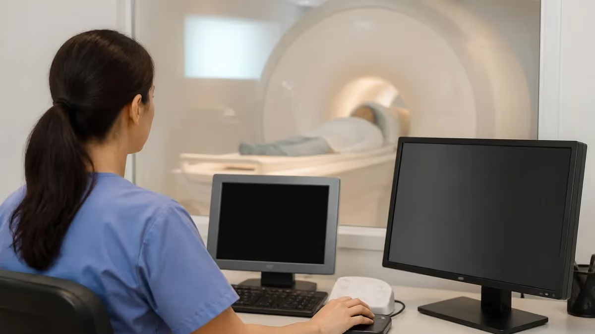
An elbow MRI is one of the most detailed musculoskeletal imaging studies in dwi mri, providing exquisite soft tissue contrast that no other modality can match. When orthopedic surgeons, sports medicine physicians, and rheumatologists need to evaluate tendon tears, ligament injuries, cartilage damage, or occult fractures, magnetic resonance imaging of the elbow becomes the test of choice. The combination of high-resolution sequences and multiplanar imaging allows radiologists to identify pathology that plain radiographs and ultrasound often miss entirely.
The elbow is a remarkably complex joint, comprising three articulations bundled into a single synovial cavity: the humeroulnar, humeroradial, and proximal radioulnar joints. Around these articulations course twelve major muscles, several critical ligaments including the ulnar and radial collateral ligaments, three major peripheral nerves, and intricate vascular structures. Imaging this density of anatomy demands meticulous protocol planning, optimal patient positioning, and a thorough understanding of how each pathology manifests on different pulse sequences.
Technologists performing an elbow MRI must balance several competing demands. The patient needs to remain perfectly still for thirty to forty-five minutes, the elbow must be positioned to maximize signal from a dedicated extremity coil, and the imaging planes must be prescribed precisely along anatomical landmarks. Even slight obliquity in slice prescription can cause the ulnar collateral ligament to appear partially torn when it is in fact intact, leading to misdiagnosis and unnecessary intervention.
Beyond technique, the diagnostic value of elbow MRI rests on the radiologist's ability to interpret subtle signal abnormalities in the context of clinical history. A throwing athlete with medial elbow pain has a different differential than a manual laborer with lateral epicondylitis or an elderly patient with insidious stiffness. The same finding, such as mild common extensor tendon thickening, may represent acute injury, chronic tendinopathy, or post-treatment scarring depending on the clinical setting and signal characteristics.
This guide walks through everything you need to know about elbow MRI, from coil selection and positioning to sequence selection, normal anatomy, common pathologies, and registry-style facts that frequently appear on certification exams. Whether you are a technologist preparing for the ARRT MRI registry, a radiology resident reading your first elbow studies, or a clinician trying to better understand the reports you receive, the information below distills the most clinically relevant points into a single resource.
We will examine the technical foundations of elbow imaging, including field strength considerations, coil selection between flexible and dedicated wrist coils, patient positioning options like the superman position versus arm at the side, and the standard sequence menu of T1, T2 fat-saturated, proton density, and STIR imaging. We will also cover indirect and direct MR arthrography, which significantly improve sensitivity for subtle ligament and cartilage injuries in throwing athletes.
By the end, you should be able to identify the structures evaluated by every plane and sequence, recognize the most common pathology patterns, and understand why certain protocols are chosen for specific clinical questions. The goal is practical, exam-ready knowledge that translates directly to better imaging and more accurate interpretation in daily practice.
Elbow MRI by the Numbers
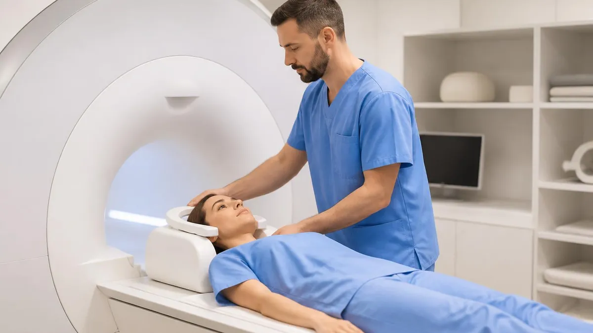
Common Indications for Elbow MRI
Throwing athletes with medial elbow pain, particularly baseball pitchers, are evaluated for UCL tears that may require Tommy John reconstruction. MRI shows partial versus complete tears and chronic thickening with greater accuracy than physical exam.
Tennis elbow and golfer's elbow involve the common extensor and flexor tendon origins respectively. MRI confirms tendinosis, partial tears, and helps grade severity when conservative treatment fails after six to twelve weeks.
Capitellar osteochondritis dissecans is common in adolescent gymnasts and throwers. MRI identifies cartilage integrity, subchondral edema, and loose body formation to guide surgical decisions between conservative care and arthroscopic intervention.
Cubital tunnel syndrome compresses the ulnar nerve along the medial elbow, while radial tunnel and pronator syndromes affect other peripheral nerves. MRI shows nerve enlargement, signal change, and mass effect from cysts or accessory muscles.
Radial head fractures, stress reactions, and bone marrow lesions often appear normal on radiographs but are obvious on MRI. The modality is also indispensable for characterizing soft tissue and bone tumors before biopsy or resection.
Successful elbow MRI begins with appropriate coil selection. Dedicated extremity coils designed specifically for the elbow or wrist provide the highest signal-to-noise ratio because they conform closely to the joint, but they limit positioning flexibility. Flexible surface coils wrap around the elbow regardless of position and are particularly useful when the patient cannot tolerate the superman position. The trade-off is slightly lower SNR, which can be partially offset by increasing the number of signal averages or using a smaller field of view.
Patient positioning is one of the most consequential decisions in elbow imaging. The superman position, where the patient lies prone with the affected arm extended overhead, places the elbow near isocenter and yields the best image quality with minimal magnetic field inhomogeneity. However, many patients find this position painful or claustrophobic, particularly those with shoulder pathology, large body habitus, or anxiety. The supine position with the arm at the side is more comfortable but moves the elbow off-isocenter, increasing susceptibility artifacts and signal drop-off.
Once positioned, the elbow should be in full extension with the palm facing upward in supination, unless the clinical question requires a specific position. Supination tightens the biceps tendon and lateral ligaments, helping evaluate distal biceps insertion at the radial tuberosity. Some institutions use the FABS view, an acronym for flexed elbow, abducted shoulder, supinated forearm, which provides a single coronal-oblique image displaying the entire distal biceps tendon from musculotendinous junction to insertion.
Field strength matters more for the elbow than for many other joints because of the small size of critical structures. The ulnar collateral ligament measures only a few millimeters thick, and partial-thickness undersurface tears at the humeral attachment can be subtle even on 3T systems. Higher field strength provides better signal and allows thinner slices, which reduces partial volume artifact. For more on the underlying physics, our explainer on how does an MRI work details the relationship between field strength, signal, and resolution.
Slice thickness for the elbow should generally fall between two and three millimeters with minimal gap, ideally zero gap when using 3D sequences. Field of view typically ranges from twelve to sixteen centimeters depending on patient size and clinical question. Matrix sizes of 256 by 256 or higher are standard, with frequency-encoded direction usually placed along the long axis of the humerus to minimize wraparound artifact from the chest wall.
Saturation bands are critical when imaging the elbow in the arm-at-the-side position. A saturation band placed over the torso eliminates respiratory motion artifact that would otherwise project across the elbow. Spatial saturation pulses also help suppress signal from arterial inflow when imaging perpendicular to vessels, reducing flow-related ghosting that can mimic pathology in nerves or tendons.
Finally, communication with the patient throughout the examination is essential. The elbow is uncomfortable in extension for prolonged periods, particularly in the superman position. Pausing briefly between sequences, adjusting padding, and providing reassurance reduces motion artifact and improves patient cooperation, which directly translates to diagnostic image quality.
MRI Practice Test Questions
Prepare for the MRI - Magnetic Resonance Imaging exam with our free practice test modules. Each quiz covers key topics to help you pass on your first try.
MRI Knowledge
MRI Exam Questions covering Knowledge. Master MRI Test concepts for certification prep.
MRI Physics
Free MRI Practice Test featuring Physics. Improve your MRI Exam score with mock test prep.
MRI Anatomy and Pathology
MRI Test Prep for MRI Anatomy and Pathology. Practice MRI Quiz questions and boost your score.
MRI Anatomy and Positioning
MRI Questions and Answers on MRI Anatomy and Positioning. Free MRI practice for exam readiness.
MRI Contrast Agents
Free MRI Quiz on MRI Contrast Agents. MRI Exam prep questions with detailed explanations.
MRI Patient Care and Positioning
MRI Practice Questions for MRI Patient Care and Positioning. Build confidence for your MRI certification exam.
Elbow MRI Pulse Sequences and Imaging Planes
Axial images are prescribed perpendicular to the long axis of the humerus through the elbow joint and proximal forearm. This plane is essential for evaluating the median, ulnar, and radial nerves as they course through their respective tunnels. The cubital tunnel containing the ulnar nerve is best assessed on axial T2 fat-saturated images, where pathologic signal change and nerve enlargement become conspicuous against the dark background of fat suppression.
The axial plane also displays the common flexor and extensor tendon origins, the annular ligament wrapping around the radial head, and the relationship between the biceps and brachialis tendons distally. Axial images extending from the distal humerus to the radial tuberosity allow complete evaluation of the distal biceps tendon as it spirals to insert on the radius, a structure frequently injured in middle-aged males during eccentric lifting.
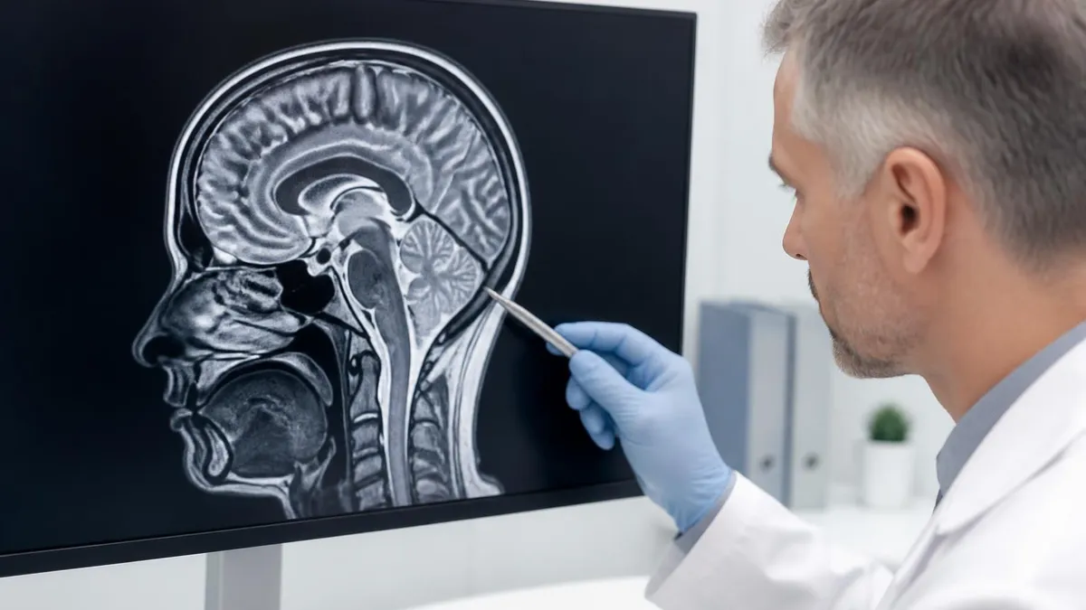
Conventional Elbow MRI vs MR Arthrography
- +Higher sensitivity for partial-thickness UCL tears, especially undersurface fiber disruption
- +Better detection of subtle chondral defects and intraarticular loose bodies
- +Improved evaluation of capsular structures distended by contrast
- +Particularly useful in throwing athletes with normal conventional MRI
- +Allows simultaneous diagnostic and therapeutic intraarticular injection
- +Distends the joint to separate adjacent structures and improve conspicuity
- −Invasive procedure requiring fluoroscopic or ultrasound guidance for needle placement
- −Small risk of infection, bleeding, or contrast reaction at injection site
- −Significantly longer total examination time including injection and transport
- −Higher cost and reimbursement complexity compared to standard MRI
- −Requires expertise for both injection technique and image interpretation
- −May overdiagnose ligament laxity in normal asymptomatic patients
Elbow MRI Pre-Scan Checklist
- ✓Screen patient for ferromagnetic implants, pacemakers, and prior orthopedic hardware
- ✓Confirm clinical question with referring provider to tailor sequences appropriately
- ✓Remove all metallic objects including watches, jewelry, and clothing with metal fasteners
- ✓Select appropriate coil based on patient size and ability to tolerate superman position
- ✓Position elbow in full extension with forearm supinated unless protocol specifies otherwise
- ✓Apply saturation bands over torso when scanning in arm-at-side position to reduce artifact
- ✓Verify isocenter placement and adjust patient position if elbow is significantly off-center
- ✓Confirm IV access patency if contrast administration is planned for the examination
- ✓Provide hearing protection and call ball before initiating any pulse sequences
- ✓Review localizer images for adequate coverage and proper anatomical orientation before proceeding
The UCL Anterior Bundle Is the Money Structure
When evaluating an elbow MRI for a throwing athlete, the anterior bundle of the ulnar collateral ligament is the single most important structure. It originates from the anteroinferior aspect of the medial epicondyle and inserts on the sublime tubercle of the ulna. Undersurface partial-thickness tears at the humeral attachment, called the T-sign on arthrography, are a classic finding in pitchers and may require surgical reconstruction.
Normal elbow anatomy on MRI begins with the osseous structures. The distal humerus consists of the medial epicondyle, lateral epicondyle, trochlea, and capitellum. The trochlea articulates with the ulnar trochlear notch, while the capitellum articulates with the radial head. Bone marrow signal should be uniform and follow the expected fat signal on T1 and fluid-sensitive sequences. Subtle marrow edema, appearing as high signal on T2 fat-saturated or STIR images and low signal on T1, may indicate stress reaction, contusion, or occult fracture.
The articular cartilage covering the trochlea, capitellum, radial head, and ulnar trochlear notch should appear smooth and intermediate signal on proton density sequences. Cartilage thickness in the elbow is thinner than in weight-bearing joints, typically measuring one to two millimeters, which demands high-resolution imaging to detect focal defects. Osteochondritis dissecans of the capitellum classically appears in adolescent athletes and shows variable signal depending on stability, with surrounding edema indicating active disease.
The medial soft tissues include the common flexor tendon origin and the ulnar collateral ligament complex. The common flexor tendon arises from the medial epicondyle and should appear uniformly low signal. Increased intratendinous signal indicates tendinosis, while a discrete defect represents partial tearing. The UCL anterior bundle runs deep to the flexor mass and should appear as a continuous low-signal band from medial epicondyle to sublime tubercle. Any waviness, thickening, or fluid signal within the ligament suggests injury.
Laterally, the common extensor tendon and lateral collateral ligament complex demonstrate similar normal appearances. The lateral ulnar collateral ligament is particularly important because its disruption causes posterolateral rotatory instability, a condition often missed on physical examination but well visualized on coronal MRI. The radial collateral ligament blends with the annular ligament wrapping around the radial head, and together these structures stabilize the radiocapitellar joint during forearm rotation.
Anteriorly, the distal biceps tendon courses from the musculotendinous junction to insert on the radial tuberosity, while the brachialis muscle inserts more medially on the ulnar tuberosity. Distal biceps ruptures, sometimes called Popeye deformity when the muscle belly retracts proximally, are well demonstrated on axial and sagittal images. The FABS view provides a single image showing the entire tendon course, which can be invaluable for surgical planning when reattachment is considered.
Posteriorly, the triceps tendon inserts on the olecranon process and the anconeus muscle lies just lateral to the triceps. Triceps ruptures are relatively rare and often associated with anabolic steroid use or systemic disease. The olecranon bursa overlying the tip of the olecranon may become inflamed in olecranon bursitis, appearing as a well-circumscribed fluid collection on T2 sequences with variable wall thickening and surrounding edema.
The three peripheral nerves crossing the elbow each have characteristic locations and pathology patterns. The ulnar nerve passes through the cubital tunnel posterior to the medial epicondyle, the median nerve travels with the brachial artery anteriorly before passing through the pronator teres, and the radial nerve divides into superficial and deep branches near the radial head. Nerve signal should be slightly higher than muscle on T2 sequences without enlargement. Hyperintensity, enlargement, or mass effect from cysts and accessory muscles indicates entrapment neuropathy.
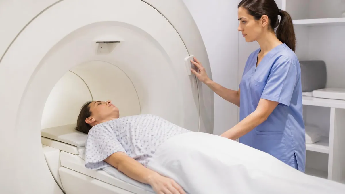
When a tendon is oriented at approximately 55 degrees to the main magnetic field, the magic angle phenomenon causes artifactually increased signal on short TE sequences like proton density. This can mimic tendinosis or partial tearing, particularly in the common extensor and flexor tendons. Always confirm suspicious findings on T2 fat-saturated sequences, where true pathology persists but magic angle effect resolves.
Reading an elbow MRI systematically prevents missed findings and reduces interpretive errors. Most experienced radiologists develop a consistent search pattern that addresses each anatomical compartment in sequence. A typical approach starts with the osseous structures, then proceeds through the medial, lateral, anterior, and posterior soft tissues, finishing with the neurovascular structures and any incidental findings in the visualized portions of the humerus, forearm, and surrounding soft tissues.
When evaluating the bones, focus on marrow signal patterns, cortical integrity, and articular surface contour. Subchondral cystic changes, edema patterns, and erosions each suggest different etiologies. Diffuse marrow edema with cortical disruption favors fracture, while a well-defined subchondral cyst with intact overlying cartilage suggests degenerative change. Comparison with prior imaging, when available, helps distinguish chronic from acute findings. For broader context on cross-modality differences, see our comparison of whats difference between mri and ct scan.
The medial elbow examination must include detailed assessment of both the common flexor tendon and the UCL anterior bundle. These two structures are intimately associated, and pathology often involves both. A common pitfall is missing a partial undersurface UCL tear in the setting of obvious flexor tendinosis because the eye is drawn to the more conspicuous finding. Always trace the UCL fiber by fiber from origin to insertion, particularly on thin-section coronal images, before concluding the ligament is intact.
On the lateral side, the common extensor tendon receives the most clinical attention because tennis elbow is so prevalent. However, the lateral ulnar collateral ligament should not be overlooked, especially in patients with prior elbow dislocation or instability symptoms. The LUCL originates from the lateral epicondyle and wraps posteriorly to insert on the supinator crest of the ulna. Its disruption may be subtle, requiring careful inspection on coronal and oblique coronal images.
Cartilage evaluation requires high-resolution proton density or T2 sequences without fat suppression to optimize contrast between cartilage and underlying bone. Focal cartilage defects, particularly on the capitellum in young athletes, may be subtle and easily missed if the slice thickness is too generous. Comparing the anterior and posterior aspects of the capitellum on consecutive sagittal images helps detect early osteochondritis dissecans before frank fragmentation occurs.
Loose bodies are best identified on the GRE or 3D sequences if available, but they can also be detected on standard T2 sequences as well-defined low-signal foci within the joint, often settling in the dependent portions of the olecranon or coronoid fossae. Multiple loose bodies suggest primary synovial osteochondromatosis, while a single fragment is more commonly post-traumatic or related to osteochondritis.
Finally, do not forget to evaluate the visualized portions of structures outside the elbow joint itself. Distal humeral lesions, proximal radial and ulnar pathology, and surrounding soft tissue masses can all be incidentally identified on elbow MRI. Reporting these findings is just as important as evaluating the primary clinical question, since unexpected findings often have significant management implications.
Practical tips for both technologists and interpreters can dramatically improve elbow MRI quality and diagnostic accuracy. The first and most important is patient preparation. Take the time before scanning to explain the procedure, the importance of stillness, and the expected duration. Patients who understand what to expect cooperate better, hold position more reliably, and produce diagnostic images on the first attempt rather than requiring repeat sequences that prolong the examination and increase the chance of further motion artifact.
Padding and immobilization deserve particular attention for elbow studies. The arm-at-side position benefits from foam cushions under the wrist and shoulder to prevent rolling, while the superman position requires careful pillow placement to support the head and minimize neck strain. Sandbags or straps placed lightly over the forearm can remind patients to remain still without causing discomfort. Comfort directly correlates with image quality in extremity imaging more than perhaps any other body part.
Sequence selection should be tailored to the clinical question rather than applied as a rigid protocol. For suspected UCL injury in a throwing athlete, thin-section coronal proton density and T2 fat-saturated sequences are essential, and MR arthrography should be considered if conventional images are equivocal. For lateral epicondylitis follow-up, axial and coronal T2 fat-saturated sequences usually suffice. For osteochondritis dissecans, sagittal sequences provide critical information about lesion stability and overlying cartilage integrity.
When interpreting elbow studies, always correlate imaging findings with the clinical history. The same anatomical finding can have different clinical significance depending on patient age, occupation, sport, and symptom duration. A small focus of common extensor tendinosis in an asymptomatic recreational tennis player is normal aging, while the same finding in a professional athlete with acute lateral elbow pain may warrant intervention. Reporting style should reflect this clinical context rather than reciting findings in isolation.
Comparison with prior imaging, when available, transforms the interpretation. A stable finding over months is much more reassuring than a new finding, even if the absolute appearance is identical. Make a habit of requesting prior studies before finalizing reports, particularly for athletes who undergo regular imaging surveillance. The interval change often guides management more than any single examination.
Consider also the role of dynamic imaging in select cases. Although standard MRI is performed in a fixed position, some institutions offer kinematic studies with the elbow imaged in flexion and extension. These can demonstrate impingement, snapping, and instability that are not appreciated on static images. While not routine, dynamic imaging fills an important diagnostic gap for patients with positional symptoms and normal conventional MRI.
Lastly, communicate clearly and proactively with referring providers. Critical findings such as complete tendon ruptures, large effusions concerning for septic arthritis, or unexpected masses should be conveyed promptly by phone in addition to the dictated report. Building this clinical communication habit improves patient care, strengthens relationships with referring services, and reinforces the radiologist's role as a consultant rather than simply an image interpreter.
MRI Questions and Answers
MRI MARS Protocol: Complete Guide to Metal Artifact Reduction Sequences for Orthopedic Imaging
How Does an MRI Work: Magnetic Resonance Imaging Explained
MRI With or Without Contrast: What to Expect
The History of MRI: From Discovery to Modern Medicine
What's the Difference Between MRI and CT Scan? A Complete Comparison Guide
About the Author

Medical Laboratory Scientist & Clinical Certification Expert
Johns Hopkins UniversityDr. Sandra Kim holds a PhD in Clinical Laboratory Science from Johns Hopkins University and is certified as a Medical Technologist (MT) and Medical Laboratory Scientist (MLS) through ASCP. With 16 years of clinical laboratory experience spanning hematology, microbiology, and molecular diagnostics, she prepares candidates for ASCP board exams, MLT, MLS, and specialist certification tests.
Join the Discussion
Connect with other students preparing for this exam. Share tips, ask questions, and get advice from people who have been there.
View discussion (8 replies)