
ARRT MRI Registry Study Guide: Complete Certification Prep for 2026 September
Master the ARRT MRI registry exam ✅ with this complete study guide covering physics, safety, protocols, and free practice questions.
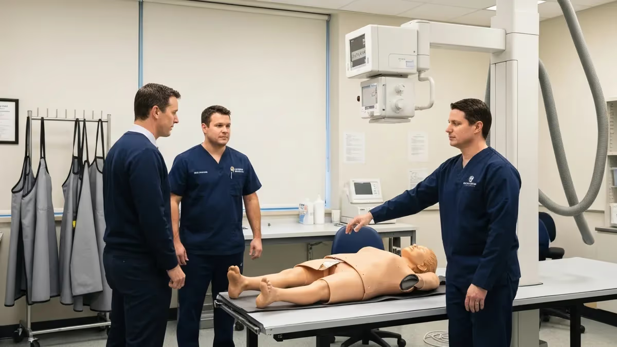
The ARRT MRI registry study guide you choose can make or break your path to MRI certification. The ARRT MRI examination is a post-primary credential designed for registered radiologic technologists who want to specialize in magnetic resonance imaging. Unlike the entry-level radiography exam, the MRI registry tests advanced knowledge of magnetic field physics, pulse sequences, patient safety protocols, and image optimization — content that demands a structured, multi-week study plan rather than a last-minute cram session. Choosing the right materials and building a repeatable daily routine are the first steps every successful candidate takes.
The MRI registry examination contains 200 questions, of which 175 are scored and 25 are unscored pilot questions embedded randomly throughout the exam. Candidates receive a maximum of 3 hours and 15 minutes to complete the test at a Pearson VUE testing center. The content outline is divided into four major categories: Patient Care, Imaging Procedures, Data Acquisition and Processing, and Physical Principles of Image Formation. Understanding the weight of each domain helps you allocate study hours where they will produce the greatest score gains rather than spreading effort evenly across topics you already know well.
Many candidates underestimate how much MRI physics departs from conventional radiography. In X-ray and CT, you are working with ionizing radiation and kilovoltage; in MRI, you are manipulating radiofrequency pulses, gradient magnetic fields, and precessing hydrogen protons. The Larmor frequency, T1 and T2 relaxation times, k-space filling strategies, and artifact recognition are all topics that require conceptual understanding, not just memorization. Building a solid physics foundation early in your study timeline will pay dividends when you encounter clinical scenario questions later, because those questions often hinge on a physics concept applied to a real patient situation.
Patient safety is another area where the ARRT MRI exam goes significantly deeper than general radiography content. The exam tests your knowledge of the four MRI safety zones (Zone I through Zone IV), ACR ferromagnetic screening guidelines, implant screening procedures, and the management of emergencies inside the MRI suite.
Thermal injury from radiofrequency energy deposition, acoustic noise hazards, and contrast agent safety — particularly the risk of nephrogenic systemic fibrosis with gadolinium-based agents — all appear on the registry. Thorough candidates treat patient safety not as a subset of the exam but as a parallel thread woven through every other content domain.
Imaging procedures make up a substantial portion of the exam and cover every major body region: brain, spine, musculoskeletal system, abdomen, pelvis, cardiovascular structures, and vascular anatomy. For each region you need to know standard patient positioning, appropriate coil selection, landmark identification, and the rationale behind sequence choices. Why do you use FLAIR for periventricular lesions? Why is STIR preferred over fat-saturated T2 for musculoskeletal edema? The exam rewards candidates who understand the clinical logic behind protocol decisions, not just those who can list sequence parameters from memory.
Data acquisition and processing includes pulse sequence design, echo time (TE), repetition time (TR), flip angle, field of view, matrix size, slice thickness, and bandwidth — parameters that interact with each other in ways that affect SNR, resolution, and scan time simultaneously. Understanding these trade-offs requires hands-on practice as well as reading. If you have access to an MRI scanner at your clinical site, spend time deliberately varying parameters and observing the results. That experiential knowledge is extremely difficult to replicate through textbook study alone and significantly reinforces theoretical concepts you encounter when reviewing an arrt mri study guide.
Committing to a realistic study timeline is perhaps the most important factor in registry success. Most candidates who pass on their first attempt report studying for 8 to 12 weeks, averaging 10 to 15 hours per week. Spread across that timeline, this means covering roughly one major content domain per two weeks, followed by two weeks of integrated practice testing and review. The sections below break down a week-by-week plan, the highest-yield content areas, and practical test-day strategies that will put you in the best possible position when you sit down at the Pearson VUE terminal.
ARRT MRI Exam by the Numbers
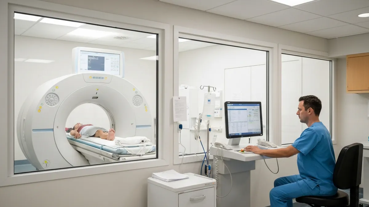
8-Week ARRT MRI Registry Study Schedule
- ▸Review magnetic field properties: static field (B0), RF pulses, gradient fields
- ▸Master T1 and T2 relaxation concepts with tissue-specific values
- ▸Study Larmor frequency and precession principles
- ▸Complete 30 physics practice questions and review all incorrect answers
- ▸Learn spin echo (SE), gradient echo (GRE), and inversion recovery sequences
- ▸Understand k-space: filling order, partial Fourier, and EPI trajectories
- ▸Study TE, TR, TI, and flip angle effects on tissue contrast
- ▸Practice identifying sequence types from tissue contrast descriptions
- ▸Memorize Zone I–IV definitions and access control requirements
- ▸Study implant screening: passive, active, and MR-conditional devices
- ▸Review gadolinium contrast agents, NSF risk factors, and screening protocols
- ▸Learn thermal bioeffects, acoustic noise limits, and peripheral nerve stimulation
- ▸Study standard brain MRI sequences: T1, T2, FLAIR, DWI, and post-contrast T1
- ▸Learn cranial nerve anatomy relevant to MRI protocol selection
- ▸Review cervical, thoracic, and lumbar spine protocols and coil positioning
- ▸Practice 40 procedure-based questions for neuro MRI
- ▸Study abdominal MRI: liver, pancreas, kidneys, and biliary system protocols
- ▸Review pelvic MRI for prostate, uterus, and rectum
- ▸Learn MSK protocols: shoulder, knee, hip, ankle, and wrist
- ▸Understand fat suppression techniques: STIR vs. fat-sat vs. Dixon
- ▸Study cardiac MRI: gating strategies, functional imaging, and viability protocols
- ▸Learn MRA techniques: time-of-flight (TOF), phase-contrast (PC), and CE-MRA
- ▸Review diffusion-weighted imaging (DWI) and apparent diffusion coefficient (ADC)
- ▸Study MR spectroscopy and perfusion imaging basics
- ▸Identify and explain all major MRI artifacts: motion, wrap-around, chemical shift, etc.
- ▸Study SNR optimization: field strength, coil selection, voxel size, and NEX/NSA
- ▸Review parallel imaging techniques: SENSE, GRAPPA, and iPAT
- ▸Practice artifact recognition with 50 image-based questions
- ▸Complete two full 175-question timed practice exams
- ▸Analyze weak areas from both exams and re-read corresponding chapters
- ▸Review all patient care topics: monitoring, contrast reactions, positioning
- ▸Final day: light review only — rest, hydrate, and confirm test center logistics
MRI physics is the domain that most candidates cite as their biggest challenge, and for good reason: the underlying science is genuinely different from anything covered in the radiography entry-level curriculum. The foundation begins with the concept of net magnetization.
When a patient is placed inside the bore of an MRI scanner, hydrogen protons in their body align with the strong static magnetic field (B0). This alignment creates a net longitudinal magnetization vector that can be manipulated by radiofrequency pulses tuned precisely to the Larmor frequency — the resonant frequency at which hydrogen protons absorb and re-emit energy at a given field strength. At 1.5 Tesla, the Larmor frequency for hydrogen is approximately 63.87 MHz; at 3 Tesla it doubles to roughly 127.74 MHz. These numbers appear on the registry exam, so memorize them.
T1 relaxation (also called longitudinal or spin-lattice relaxation) describes how quickly protons return their energy to the surrounding molecular lattice and re-establish their longitudinal magnetization after an RF pulse. Fat has a very short T1 time and therefore appears bright on T1-weighted images. Cerebrospinal fluid has a very long T1 time and appears dark. Understanding these tissue-specific values lets you predict image contrast without having to memorize every possible sequence parameter combination, which is exactly the kind of flexible reasoning the exam demands in clinical scenario questions.
T2 relaxation (transverse or spin-spin relaxation) describes how quickly the transverse magnetization decays due to interactions between neighboring protons. Water has a very long T2 time and appears bright on T2-weighted images — which is why T2-weighted sequences are preferred for detecting edema, inflammation, and most pathological processes that increase tissue water content.
T2* (T2-star) relaxation adds the effects of local magnetic field inhomogeneities to the basic T2 decay and is the contrast mechanism exploited by gradient echo sequences used in functional MRI (fMRI) and susceptibility-weighted imaging (SWI). Hemorrhage, calcium, and iron produce T2* shortening and therefore appear dark on SWI sequences.
Inversion recovery sequences are a high-yield physics topic because they allow selective suppression of specific tissues based on their T1 relaxation times. Short TI Inversion Recovery (STIR) suppresses fat by applying the inversion pulse and then waiting until fat's longitudinal magnetization crosses the null point before collecting data.
Fluid-Attenuated Inversion Recovery (FLAIR) uses a longer inversion time to null free water (CSF), making periventricular lesions, cortical lesions, and subarachnoid hemorrhage much more conspicuous than on standard T2-weighted images. The ARRT registry frequently presents clinical scenarios asking which sequence is most appropriate for a given clinical question, and inversion recovery sequences appear prominently in those questions.
MRI safety demands equal attention. The ACR's Zone classification system defines Zone I as public areas outside the MRI environment, Zone II as the transition space between public and restricted areas, Zone III as the area with controlled access that houses the scanner room entrance, and Zone IV as the MRI scanner room itself — the most restricted zone where only screened personnel and patients may enter.
Ferromagnetic projectile incidents (the so-called missile effect) occur when ferromagnetic objects are inadvertently brought into Zone IV and accelerate toward the bore due to the strong attractive force of B0. These incidents have caused serious injuries and deaths, making ferromagnetic screening one of the most critical competencies tested on the registry.
Gadolinium-based contrast agents (GBCAs) are another patient safety topic with significant clinical depth on the registry. There are two major categories: linear agents (which are less stable and associated with higher gadolinium retention and the risk of nephrogenic systemic fibrosis in patients with severely reduced renal function) and macrocyclic agents (which are more stable and associated with lower retention). Current ACR guidelines recommend screening all patients for renal function before administering GBCAs, particularly for patients with chronic kidney disease, acute kidney injury, or who are on dialysis.
The registry tests not only when to withhold contrast but also how to manage acute contrast reactions, including the distinction between allergic-like reactions and physiologic reactions and the appropriate clinical response to each.
Artifacts represent a crossroads between physics and clinical practice. Motion artifact — the most common artifact encountered in clinical MRI — appears as ghosting or blurring along the phase-encoding direction and results from patient movement or physiologic motion (cardiac pulsation, respiratory motion, peristalsis) during data acquisition.
Remedies include breath-hold techniques, respiratory triggering or gating, cardiac gating, faster pulse sequences, and patient coaching. Wrap-around (aliasing) artifact occurs when the field of view is smaller than the anatomy being imaged, causing structures outside the FOV to appear folded back onto the image. It can be corrected by increasing the FOV, using oversampling in the phase direction, or selecting a saturation band over the offending anatomy.
ARRT MRI Exam Content Domain Breakdown
Physical Principles of Image Formation accounts for approximately 30% of the ARRT MRI registry exam, making it the single largest content domain by weight. Topics include magnetic field properties, RF excitation, relaxation mechanisms (T1, T2, T2*), pulse sequence design, k-space theory, Fourier transformation, gradient function (slice selection, frequency encoding, phase encoding), and SNR optimization. Candidates must understand not only how each parameter works in isolation but also how changing one parameter — such as reducing slice thickness — affects resolution, SNR, and scan time simultaneously.
Artifact recognition and correction is embedded within the physical principles domain and constitutes some of the most frequently tested material on the registry. The major artifacts — motion, wrap-around, chemical shift (first and second order), susceptibility, zipper, truncation (Gibbs ringing), and cross-talk — each have specific causes and specific remedies. For example, chemical shift artifact of the second kind (India ink artifact) occurs at the boundary between fat and water at certain TE values in gradient echo sequences; it is corrected by adjusting TE to an in-phase value. Mastery of artifact cause-and-effect pairs is essential for scoring well in this domain.

Self-Study vs. Structured Review Course: Which Is Right for You?
- +Self-study allows complete schedule flexibility — ideal for technologists with variable shift work
- +Textbook-based review lets you spend extra time on physics, which is the most heavily weighted domain
- +Free and low-cost resources (practice questions, ARRT content outline, ACR guidelines) cover most high-yield content
- +Learning at your own pace reduces burnout compared to compressed live review courses
- +Combining self-study with a question bank builds active recall, which improves long-term retention
- +You can customize your study plan to target your personal weak areas identified through diagnostic practice tests
- −Self-study requires strong self-discipline — many candidates underestimate the time commitment and fall behind
- −Without a structured course, it is easy to over-study low-yield topics and neglect high-yield areas
- −Physics concepts can be genuinely confusing without an instructor to clarify abstract principles in real time
- −Quality of free online resources varies widely — some contain outdated or inaccurate information
- −Lack of peer accountability means study sessions are easier to skip, especially during long study timelines
- −Timed practice exams under realistic conditions are harder to replicate without a formal course platform
ARRT MRI Registry Exam Preparation Checklist
- ✓Download the official ARRT MRI Content Outline from arrt.org and highlight every topic you feel uncertain about.
- ✓Schedule your Pearson VUE exam date at least 6 weeks out so you have a firm deadline to study toward.
- ✓Purchase or borrow a dedicated MRI review textbook covering physics, sequences, and clinical protocols.
- ✓Set up a question bank account and complete at least 500 practice questions before exam day.
- ✓Build a weekly study calendar that blocks 2 hours per day, five days per week, for the full 8-week period.
- ✓Create a flashcard deck for the Larmor frequency values at 1.5T and 3T, T1/T2 tissue values, and artifact causes.
- ✓Review the ACR MRI Safety Manual and memorize the four-zone classification system with access requirements.
- ✓Practice identifying artifacts on sample images and writing out the correction strategy for each artifact type.
- ✓Complete two full-length 175-question timed practice exams in the final two weeks of your study plan.
- ✓Confirm your testing center location, parking, valid photo ID requirements, and center-specific check-in rules one week before exam day.
The Physical Principles Domain Is Your Highest-Leverage Study Target
Physical Principles of Image Formation carries approximately 30% of the total exam weight, making it the single most influential content domain on the ARRT MRI registry. Candidates who score strongly in physics consistently outperform those who focus only on procedures. Spend at least 3 full weeks of your study plan on physics before pivoting to clinical protocols — your score will reflect the investment.
High-yield imaging procedures for the ARRT MRI registry begin with neurologic MRI, and specifically with brain protocols. A standard brain MRI at most institutions includes axial T1, axial T2, axial FLAIR, axial DWI with ADC map, and post-contrast axial and sagittal T1 sequences. The registry tests why each sequence contributes uniquely to the diagnostic picture.
FLAIR suppresses CSF signal, making it the preferred sequence for detecting periventricular white matter lesions in multiple sclerosis, cortical lesions that would otherwise be obscured by adjacent bright CSF, and leptomeningeal enhancement. DWI detects acute ischemic stroke within minutes of onset — far earlier than T2-weighted sequences — because cytotoxic edema restricts water diffusion and produces bright signal on DWI with corresponding low ADC values.
Spine MRI is another high-yield procedural area, particularly given its prevalence in clinical practice.
The ARRT MRI exam tests appropriate coil selection (posterior phased array for cervical and lumbar spine), standard plane selection (sagittal T1 and T2 are always acquired, with axial sequences added through areas of pathology), and the clinical indications that differentiate lumbar disc herniation (which compresses nerve roots and is typically evaluated with T2 axial sequences through the disc levels of interest) from vertebral marrow pathology (where T1 signal loss indicates marrow replacement and STIR increases sensitivity for edema). Sagittal STIR is the sequence of choice when spinal cord contusion, vertebral fracture, or metastatic disease is suspected.
Musculoskeletal MRI is extensive and covers all major joints.
For knee MRI — one of the most commonly performed MRI examinations in clinical practice — candidates should know the standard protocol: coronal PD fat-sat (for menisci and collateral ligaments), sagittal PD fat-sat (for cruciate ligaments and cartilage), and axial PD fat-sat (for the patellofemoral compartment). STIR is preferred over fat-saturated T2 in the setting of suspected bone marrow edema, stress fracture, or soft tissue inflammation because STIR is more robust to field inhomogeneities — particularly at field strengths below 1.5T and at anatomic regions with inherent B0 heterogeneity such as the shoulder and ankle.
Shoulder MRI offers another instructive example of protocol rationale that the registry frequently tests. For rotator cuff evaluation, coronal oblique T2 fat-sat sequences oriented parallel to the supraspinatus tendon are the workhorse, providing high conspicuity for tendon tears, tendinosis, and subacromial bursal fluid.
MR arthrography — in which diluted gadolinium is injected directly into the glenohumeral joint under fluoroscopic guidance before MRI — is the gold standard for evaluating labral pathology (Bankart lesions, SLAP tears) because the joint distension separates structures that otherwise contact each other and are indistinguishable on conventional MRI. The registry may ask you to identify which examination type is most appropriate given a specific clinical scenario.
Abdominal and pelvic MRI protocols require knowledge of liver-specific contrast agents (hepatobiliary agents such as gadoxetate disodium, which is taken up by functional hepatocytes and excreted into bile, providing both dynamic vascular phases and a hepatobiliary phase that improves detection of hepatocellular carcinoma), breath-hold technique requirements, and the role of chemical shift imaging (in-phase and opposed-phase GRE sequences) in characterizing adrenal adenomas and hepatic steatosis. Fat-containing lesions lose signal on opposed-phase images relative to in-phase images due to intracellular fat — this is a diagnostic criterion for adrenal adenoma that the registry tests in clinical scenario format.
Cardiovascular MRI is among the most technically demanding sections of the exam and has grown in emphasis on recent registry content outlines. Cardiac MRI requires prospective or retrospective ECG gating to freeze cardiac motion. Cine sequences using steady-state free precession (SSFP, also marketed as TrueFISP, FIESTA, or BFFE depending on the manufacturer) provide high blood-myocardium contrast without contrast injection and are used to assess ventricular volumes, ejection fraction, and wall motion abnormalities.
Late gadolinium enhancement (LGE) imaging, acquired 10 to 15 minutes after contrast injection, shows areas of myocardial fibrosis or infarction as bright regions because gadolinium washes out of normal myocardium but accumulates in areas of interstitial fibrosis where cell membranes are disrupted.
MR angiography techniques round out the high-yield procedural content. Time-of-flight (TOF) MRA is based on flow-related enhancement: unsaturated blood flowing into a saturated imaging slab appears bright because it has not yet experienced RF saturation. It works best for slow-flow vessels with flow perpendicular to the imaging plane and is the standard technique for intracranial MRA to evaluate for aneurysms and vascular malformations.
Phase-contrast (PC) MRA encodes velocity as a phase shift and can be used not only to depict vessels but also to quantify blood flow velocity and volume — a capability exploited in cardiac MRI for measuring cardiac output and in neuroradiology for CSF flow studies in the evaluation of normal pressure hydrocephalus.
To sit for the ARRT MRI registry examination, you must hold a current, unrestricted primary ARRT credential (typically Radiography, Nuclear Medicine, or Radiation Therapy) and meet the MRI-specific educational and clinical experience requirements. As of 2026, the structured education requirement includes didactic MRI physics and clinical MRI experience documented through the ARRT competency checklist. Verify your eligibility status on arrt.org before scheduling your Pearson VUE appointment, as eligibility lapses can delay your exam date significantly.
Building an effective practice test strategy is as important as content mastery in your final preparation phase. The research on exam performance consistently shows that active recall through practice testing produces stronger long-term retention than passive re-reading of notes or textbooks.
This principle — known as the testing effect or retrieval practice effect — means that spending 40% of your study time answering practice questions is more efficient than spending 100% of your time reading, even when reading feels more comfortable and productive. Structure your final four weeks so that practice question sessions outnumber reading sessions, and treat every incorrect answer as a learning opportunity rather than a failure.
When you review wrong answers, don't just read the correct answer and move on. Instead, write out a brief explanation of why the correct answer is right and why each distractor is wrong. This process forces you to engage with the underlying concept at a deeper level than simple recognition, which is exactly the cognitive depth required to answer novel questions on exam day.
For physics questions in particular, trace the causal chain: what property of the tissue, sequence, or parameter caused the described outcome? If you cannot answer that question in your own words, you have not yet mastered the concept and should return to your review materials before attempting more questions in that category.
Simulating real exam conditions during your final practice tests is critical. The ARRT MRI exam is 3 hours and 15 minutes long, and cognitive fatigue is a genuine performance variable — especially for questions in the final third of the exam. Practice with a strict timer, no interruptions, and no access to reference materials.
Take a 5-minute break at the midpoint if your practice platform allows it. After the simulated exam, complete a detailed performance review: identify which content domains had the highest error rates, which question types (application vs. recall) caused the most difficulty, and whether your pacing was adequate or whether you rushed the final 30 questions due to time pressure.
Time management during the real exam requires a deliberate approach. With 200 questions and 195 minutes of testing time, you have slightly less than one minute per question on average. Most candidates find that straightforward recall questions take 20 to 30 seconds, leaving extra time for complex clinical scenario questions that require careful reasoning.
A practical strategy is to move through the exam at a steady pace, flagging any question you are unsure about, and returning to flagged questions after completing the initial pass. Pearson VUE's testing interface allows question flagging and easy navigation between questions, so use this feature rather than spending disproportionate time on any single question during your first pass.
The morning of your exam, prioritize sleep and nutrition over last-minute studying. Attempting to learn new material the night before the exam typically backfires because working memory is impaired by anxiety and fatigue, making new information difficult to consolidate.
A light review of your flashcards for key formulas and values (Larmor frequencies, T1/T2 tissue values, zone definitions) is appropriate, but avoid reading entire chapters or watching lengthy video lectures. Arrive at the Pearson VUE testing center at least 30 minutes early with your valid government-issued photo ID, and take a few minutes in the car or lobby to use deliberate breathing techniques to reduce test anxiety before check-in.
Reviewing an arrt mri study guide that consolidates MRI physics, safety, and procedures into a single organized reference is one of the most efficient preparation strategies available to candidates with limited study time.
A well-structured review text allows you to move through content domains systematically, cross-reference related topics (for example, how artifact type connects to specific sequence parameters), and identify gaps in your knowledge before they show up as wrong answers on the actual registry. Supplement any review text with up-to-date ARRT content outline guidance, since the weighted distribution of exam questions shifts slightly each year as new clinical techniques become standard practice.
If you do not pass on your first attempt, understand that the ARRT allows up to three attempts per year, and that each retest provides a score report identifying content domains where performance was below the passing threshold. Use those domain scores strategically: focus your remediation study on the lowest-performing areas rather than repeating a general review of all material.
Candidates who use their score report as a targeted study guide consistently perform better on their second attempt than those who simply repeat the same broad preparation strategy. The ARRT MRI credential is a meaningful career milestone — persistent, strategic preparation is the path to earning it.
Practical test-day strategies begin long before you walk through the Pearson VUE door. In the final week before your exam, shift your schedule to match the time of day when your exam is booked. If you are scheduled for a morning slot, make sure you have been waking up and studying in the morning throughout that final week so that your cognitive performance peaks at exam time rather than mid-afternoon when your body expects to be active.
Sleep quality in the week before the exam matters more than the single night immediately before — cumulative sleep debt suppresses working memory, attention, and processing speed in ways that a single good night cannot fully reverse.
During the exam itself, read every question stem completely before looking at the answer choices. The ARRT MRI registry uses clinical scenario questions that embed the critical information in the body of the question rather than in the answer choices. Candidates who jump to the answers before finishing the stem frequently miss a qualifier — such as a patient contraindication or a specific imaging finding — that changes the correct answer entirely.
After reading the stem, predict what you believe the answer should be before revealing the choices; if your prediction matches one of the options, that choice deserves serious consideration even if a more elaborate option seems appealing.
Process of elimination is your most powerful tool for questions where you are not immediately certain of the correct answer. On a four-option multiple-choice exam, eliminating even one clearly wrong answer raises your probability of guessing correctly from 25% to 33%. Eliminating two wrong answers brings it to 50%. The ARRT MRI registry does not penalize for incorrect answers (there is no penalty for guessing), so you should always select an answer for every question rather than leaving items blank, even if you are guessing.
On questions where you have eliminated two options, your educated guess has a 50-50 chance of being correct — meaningful odds when accumulated across several uncertain questions.
After you submit your exam and receive your pass/fail result, take time to reflect on the preparation process regardless of the outcome. What study resources were most effective? Where did you lose the most time during study sessions? Were there content areas where your clinical experience provided an advantage, and areas where book knowledge alone was insufficient?
These reflections will improve your preparation process if you need a second attempt, and they may also point toward continuing education opportunities that will make you a more proficient MRI technologist in clinical practice — which is, ultimately, the purpose the ARRT registry credential is designed to serve.
Staying current with MRI technology advances is a professional obligation that extends beyond the registry exam itself. Field strength has continued to increase, with 7 Tesla systems now FDA-approved for specific clinical indications, and ultra-low field portable MRI systems are entering clinical use for point-of-care applications. Artificial intelligence is being integrated into MRI workflows for scan planning, artifact reduction, image reconstruction, and diagnostic support.
The ARRT CE requirements for MRI credential maintenance ensure that certified technologists remain current with these advances, but actively engaging with continuing education — attending ISMRM or ASNR conferences, reading Radiological Society of North America publications, or completing online CE modules — accelerates professional development well beyond the minimum CE threshold.
Building relationships with experienced MRI technologists and radiologists at your clinical site is one of the highest-value investments you can make during your preparation period. Asking a senior technologist to explain the rationale behind protocol decisions — why a sequence was added, why a parameter was changed for a specific patient — converts abstract textbook knowledge into applied clinical reasoning.
Similarly, reviewing interpreted MRI cases with a radiologist and asking about the imaging findings that drove the interpretation builds the visual pattern recognition skills that support image-based questions on the registry exam. No textbook can fully replicate the learning that comes from direct clinical mentorship.
The ARRT MRI credential opens doors to higher-paying MRI-focused positions, travel MRI contracts, lead technologist roles, and clinical educator positions that are not available to general radiographers without the post-primary credential. According to industry salary surveys, MRI technologists with their ARRT MRI registry certification earn a median annual salary roughly $8,000 to $12,000 higher than non-credentialed technologists in comparable markets. The credential also signals to employers that you have achieved a verified, standardized level of competence — a meaningful differentiator in competitive hiring markets in major metropolitan areas where multiple credentialed candidates compete for the same positions.
ARRT Questions and Answers

Mosby ARRT Review: The Complete Study Guide for Radiologic Technology Certification

ARRT Practice Test PDF (Free Printable 2026)

Is the ARRT Exam Multiple Choice? Complete ARRT Exam Format Guide 2026

How to Report CE Credits to ARRT: Complete Step-by-Step Guide

ARRT Practice Test 2026: Free Questions & Full Exam Prep Guide
About the Author

Medical Laboratory Scientist & Clinical Certification Expert
Johns Hopkins UniversityDr. Sandra Kim holds a PhD in Clinical Laboratory Science from Johns Hopkins University and is certified as a Medical Technologist (MT) and Medical Laboratory Scientist (MLS) through ASCP. With 16 years of clinical laboratory experience spanning hematology, microbiology, and molecular diagnostics, she prepares candidates for ASCP board exams, MLT, MLS, and specialist certification tests.