Meniscus Tear MRI: Imaging Findings, Grades, and What Patients Should Know
Understand meniscus tear MRI findings, grading, signal patterns, and report language so you can prepare for your scan with confidence.
A meniscus tear is one of the most common reasons a knee MRI gets ordered, and the imaging report often reads like a different language. You hear words like grade 3 signal, bucket-handle, radial, parrot-beak, and root tear, and you start wondering whether your knee will ever feel normal again. The truth is calmer than it sounds. Most meniscal abnormalities seen on MRI are stable, manageable, and well understood by orthopedic teams who read these scans every day.
This guide walks through what a meniscus tear MRI actually shows, how radiologists grade the signal, what each tear pattern means, and how providers translate the imaging into a treatment plan. You will not be quizzed on Latin anatomy. Instead, you will get a plain-language tour of the knee through the eyes of the scanner, with enough technical depth to follow the conversation when your orthopedist points at a screen.
Knee MRIs are remarkably accurate for meniscal pathology. Reported sensitivity for medial meniscus tears typically lands between 90 and 95 percent, while specificity reaches the mid 80s and higher for lateral tears. That accuracy depends on a clean scan, proper sequences, and an experienced reader. When all three line up, the report you receive is one of the most useful pieces of diagnostic information in sports medicine.
If you are preparing for your scan, recovering from a knee injury, or simply trying to make sense of a recent radiology report, the next sections will help you decode it section by section. Anatomy first, then signal grading, then tear morphology, then what shows up on the report, and finally what comes next clinically.
Meniscus Tear MRI at a Glance
Knee Anatomy as the MRI Sees It
Before you can interpret meniscus findings, it helps to picture what the scanner is actually looking at. The knee houses two C-shaped fibrocartilage discs sitting between the femur above and the tibia below. The medial meniscus is the larger, more crescent-shaped disc on the inside of the knee, attached more firmly to the joint capsule. The lateral meniscus on the outside is rounder, more mobile, and slightly less prone to certain tear patterns. Both menisci absorb shock, distribute weight across the joint, and keep articular cartilage from grinding bone on bone.
On MRI, healthy menisci appear uniformly dark on standard sequences because their dense collagen weave does not hold much free water. When you see anything brighter than that solid black wedge, the radiologist starts paying attention. The brightness, where it sits within the meniscus, and whether it reaches a surface determine whether it is a degenerative change, an intra-substance signal, or a true tear.
Each meniscus has three zones described as the anterior horn, body, and posterior horn. The posterior horn of the medial meniscus is the most common site of tears, particularly degenerative ones in middle-aged adults. The lateral meniscus tends to tear acutely in younger athletes during pivot injuries, and root tears of either meniscus carry their own clinical weight because they functionally behave like a complete meniscectomy.

The posterior horn of the medial meniscus bears the most load during knee flexion and rotation. Repeated micro-stress at this site leads to collagen disruption, which is why most degenerative tears in adults over 40 show up here. Acute tears in younger patients often follow a twisting injury with the foot planted, again loading the posterior horn under shear.
MRI Signal Grading: The Foundation of the Report
Radiologists describe meniscal abnormalities using a three-grade signal system that has been standard for decades. Each grade reflects how bright the signal is on T2 or proton density fat-suppressed sequences and whether it touches the articular surface of the meniscus.
Grade 1 signal appears as a small, globular focus of increased intensity within the substance of the meniscus that does not reach any surface. This represents early mucoid degeneration and is rarely symptomatic on its own. Many radiology reports still mention it, especially in adults over 35, but it does not equate to a tear.
Grade 2 signal is linear, often horizontal, and stays internal without reaching the articular surface. It indicates more extensive degeneration and sometimes a partial intra-substance disruption. Like grade 1, it is not classified as a tear because the meniscus surface remains intact, but it does signal aging cartilage that may progress.
Grade 3 signal is the radiologist's threshold for calling a true tear. The increased signal extends to either the superior or inferior articular surface of the meniscus. Once contrast reaches that surface, the tear is considered communicating and biomechanically significant. The report will then specify the pattern, location, and any associated displaced fragments.
Some institutions also use a grade 2A or grade 2B refinement to flag signal that nearly reaches the surface but cannot be conclusively traced. This nuance often prompts follow-up imaging or a clinical correlation note.
Meniscal Tear Patterns You Will See on MRI
Tear runs parallel to the tibial plateau, separating the meniscus into upper and lower halves like the layers of a pancake. Common in degenerative knees of patients over 40 and often associated with parameniscal cysts on the lateral side. Symptoms can include mild swelling, joint line tenderness, and occasional clicking with deep flexion. Conservative care often manages these well.
Perpendicular tear extending from the free inner edge outward toward the capsule like a slice through a pie crust. Disrupts circumferential collagen fibers and the meniscus ability to bear hoop stress, leading to early extrusion. Even small radial tears matter clinically because they accelerate cartilage breakdown if untreated, particularly when they extend into the meniscal root region.
Tear runs along the long axis of the meniscus, parallel to the curve of the joint line. Often acute and traumatic in younger patients after a twisting injury with the foot planted. Can progress into a bucket-handle if the inner fragment displaces toward the notch. Peripheral longitudinal tears in the red zone have good repair potential.
Displaced longitudinal tear where the inner fragment flips into the intercondylar notch resembling a bucket handle pulled out. Classic absent bow-tie sign and double PCL sign on coronal and sagittal images. Often locks the knee mechanically and prevents full extension. Surgical repair or partial meniscectomy is usually required.
A piece of meniscus tissue partially separated and folded over itself like a hinged trap door. Causes mechanical symptoms like catching and can mimic loose body sensations during activity. Patients often describe a feeling that something is moving inside the knee. Arthroscopic trimming of the unstable flap usually resolves symptoms.
Avulsion at the meniscal attachment to the tibial plateau where the meniscus anchors firmly to bone. Functionally equivalent to total meniscectomy because hoop tension is lost across the entire meniscus. Strongly linked to rapid cartilage breakdown if untreated, and early repair is increasingly favored for younger patients.
Reading the MRI Report Section by Section
Most knee MRI reports follow a standardized template that walks structure by structure through the joint. Knowing the order helps you focus on the meniscus paragraph rather than getting lost in everything else.
The technique section lists scanner field strength, typically 1.5 Tesla or 3 Tesla, and the pulse sequences used. You will see acronyms like PD, PDFS, T2, STIR, and sometimes T1. PD fat-suppressed sequences are the workhorse for meniscal assessment because they balance contrast resolution with water suppression. A 3T scan does not always outperform 1.5T for meniscal tears, but it does improve cartilage detail.
The findings section starts with bone and marrow, then ligaments, then menisci, then cartilage, and ends with extra-articular structures. The meniscus paragraph identifies signal abnormalities by location (anterior horn, body, posterior horn, root), grade, and tear morphology if present. A typical line might read: "Grade 3 oblique signal extending to the inferior articular surface of the posterior horn of the medial meniscus, consistent with horizontal cleavage tear."
The impression section condenses everything into a numbered list of clinically actionable findings. This is the section your orthopedist will read first. If you only have time to digest one part of the report, start at the impression and work backward to the detail.
Common MRI Sequences for Meniscus Imaging
Proton density with fat suppression is the primary workhorse sequence for meniscal evaluation. It shows fluid bright, fat dark, and meniscal tears as bright signal against the dark fibrocartilage background. Used in coronal, sagittal, and axial planes to ensure tears are not missed at any orientation. Slice thickness typically ranges from 3 to 4 millimeters for optimal meniscal detail.
Special MRI Signs That Confirm Specific Tear Types
Beyond signal grading, radiologists rely on classic imaging signs that essentially serve as quick visual confirmations of specific tear patterns. These signs are pattern-recognition shortcuts built from decades of correlation between MRI and arthroscopy.
The absent bow-tie sign refers to a bucket-handle tear. Normally, two sequential sagittal images through the body of the meniscus show a bow-tie shape of dark meniscal tissue. If fewer than two consecutive bow-ties appear, a longitudinal fragment has likely displaced into the notch.
The double PCL sign appears on a sagittal image when a displaced bucket-handle fragment lies parallel to and just anterior to the posterior cruciate ligament, creating the illusion of two PCLs. This is one of the most reliable secondary signs for a flipped fragment.
The ghost meniscus sign describes a section of meniscus that becomes nearly invisible on PDFS sequences because of complete signal alteration from a complex tear or absent tissue after fragment displacement. It is subtle but meaningful.
The cleft sign points to a radial tear, where a vertical bright line cuts across the dark meniscus from inner to outer edge. Even small radial tears matter clinically because they disrupt hoop stress and accelerate cartilage damage.
Posterior root tears of the medial meniscus often appear as a small ghost area at the attachment site with associated meniscal extrusion of 3 mm or more on coronal images. They behave biomechanically like a total meniscectomy and can lead to rapid joint space narrowing. If your report mentions extrusion or root involvement, ask whether surgical repair is being considered.
Acute vs. Degenerative: How MRI Tells Them Apart
One of the most useful distinctions an MRI provides is whether a tear looks acute, chronic, or somewhere in between. The radiologist looks at edge sharpness, surrounding edema, joint effusion, and bone marrow signal to make this call.
Acute tears typically show clean, sharp edges with bright signal, surrounding marrow edema near the joint line, and a moderate-to-large joint effusion. The patient often has a clear injury event and signs of acute synovitis. These tears are more likely to be candidates for surgical repair, especially if they sit in the vascular outer third.
Degenerative tears develop over time, usually in adults over 40. The signal is often horizontal, the edges look irregular and softened, and surrounding edema is minimal. There may be coexisting cartilage thinning, subchondral cysts, or osteophyte formation. Surgical repair offers little benefit in this setting, and conservative management with physical therapy usually outperforms arthroscopic debridement.
The vascular zones of the meniscus also matter. The outer one-third is called the red zone because it has blood supply and can heal. The middle third (red-white zone) heals less reliably. The inner third (white zone) has essentially no blood supply and rarely heals, even with surgical repair. Your MRI report will often note tear location relative to these zones because it directly influences whether repair is realistic.
What to Bring to Your Knee MRI Appointment
- ✓Photo ID and insurance card or referral paperwork
- ✓Prior knee imaging on disc or hospital network access
- ✓Complete list of metal implants including stents pacemakers or aneurysm clips
- ✓Comfortable clothing without zippers buttons or metal fasteners on the lower body
- ✓List of recent knee injuries with specific dates and mechanism of injury
- ✓Names and doses of medications especially blood thinners or pain medications
- ✓Pain or symptom diary if your clinician requested one before the scan
- ✓Questions about contrast claustrophobia or anticipated scan duration
- ✓Driver if you plan to take anti-anxiety medication before the scan
- ✓Snack and water for after the scan particularly if you skipped a meal
How MRI Influences Treatment Decisions
An MRI report is only useful if it changes the plan, and meniscal imaging does exactly that. The combination of tear type, location, vascular zone, patient age, activity level, and associated cartilage damage drives the conversation about conservative care versus surgery.
Conservative management is usually first-line for small, stable, degenerative, or low-symptom tears. This includes activity modification, structured physical therapy focused on quadriceps strengthening, NSAIDs when appropriate, and sometimes a corticosteroid or hyaluronic acid injection. Many patients return to full activity within 6 to 12 weeks.
Arthroscopic meniscectomy removes the torn portion and smooths the remaining meniscus. It is reserved for mechanical symptoms like locking or persistent pain after a full conservative trial. Studies have repeatedly shown that meniscectomy does not improve outcomes for degenerative tears compared with physical therapy in middle-aged adults without locking.
Arthroscopic meniscal repair sutures the torn edges together and is preferred in younger patients with peripheral (red zone) longitudinal or root tears. Recovery is longer (often 4 to 6 months before unrestricted activity), but preserving meniscal tissue protects cartilage and reduces the long-term risk of osteoarthritis.
Meniscal transplantation is a more specialized option for younger patients who have lost most of one meniscus. MRI helps screen candidates by quantifying remaining tissue, cartilage status, and alignment.
MRI for Meniscal Injury: Strengths and Limits
- +Non-invasive with no ionizing radiation exposure to the body
- +High sensitivity and specificity for medial meniscus tears in trained hands
- +Identifies associated ligament cartilage and bone injuries in the same scan
- +Differentiates acute from degenerative tear patterns with high reliability
- +Helps surgeons plan the arthroscopic approach and predict findings in advance
- +Allows comparison over time when prior scans are available for review
- −Lower accuracy for lateral meniscus tears in some published series
- −Cannot always distinguish stable from unstable tears without arthroscopy
- −False positives common in older patients with degenerative signal changes
- −Claustrophobia and motion artifact can reduce scan quality significantly
- −Some metal implants restrict access or limit available scan field strength
- −Cost and access vary widely depending on insurance and geographic region
Questions to Ask After You Receive Your Report
Reading your own MRI report can feel overwhelming. A short list of focused questions keeps the conversation with your provider productive and helps you leave the visit with a clear plan instead of more anxiety.
First, confirm the diagnosis: is this a true grade 3 tear or grade 2 degeneration? The distinction changes treatment dramatically. Second, ask about location and zone, because peripheral tears have repair potential while central tears generally do not. Third, ask whether the tear is acute or chronic in radiographic appearance, since this affects timeline and prognosis.
You also want to clarify what else the scan showed. Many knee MRIs reveal incidental findings like Baker cysts, mild cartilage fibrillation, or subchondral cysts that are not the source of pain but can distract from the main diagnosis. Ask your provider to rank findings by clinical relevance.
Finally, talk about timing. If conservative care is the plan, what does the next 6 to 12 weeks look like, and what milestone would prompt a return visit or surgical consultation? If surgery is on the table, ask about repair versus partial meniscectomy, recovery timelines, and any rehabilitation requirements before scheduling.
MRI Questions and Answers
What Happens During the Scan Itself
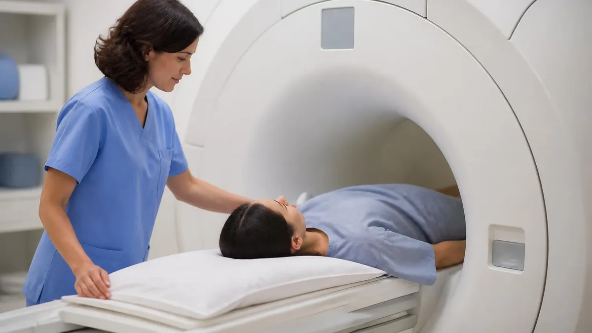
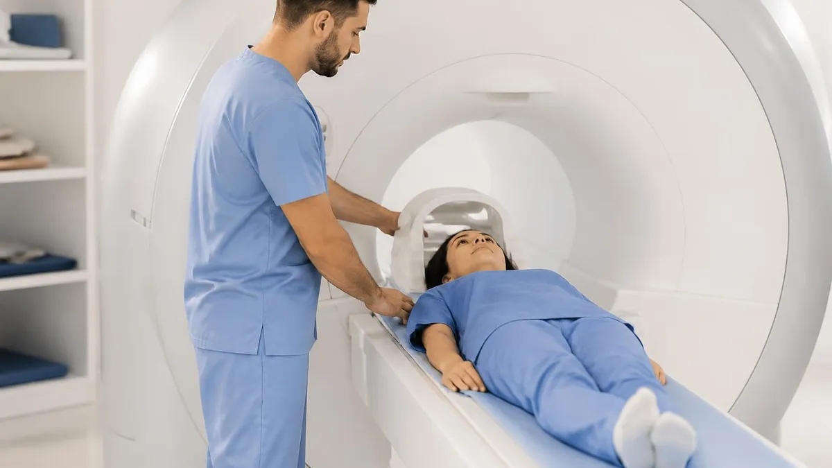
If you have never had a knee MRI, the actual experience tends to be far less dramatic than people fear. You change into a gown or scrub pants, remove anything metal, and lie down on a padded table. A small coil device, which is just a specialized antenna, wraps gently around the knee. The table slides into the bore, but for a knee scan only your lower body enters the tube. Most patients can keep their head outside the magnet, which solves the majority of claustrophobia concerns before they start.
The scanner produces loud knocking and buzzing sounds as different sequences run. You receive earplugs or headphones, and many centers play music. Each sequence lasts two to six minutes, and the technologist will remind you to hold still through the intercom. Holding the leg still is more important than holding your breath. Tiny movements blur the meniscus, which is exactly the structure being studied. Most patients finish the entire exam in under half an hour and walk out without any restrictions or recovery time.
Putting Your Meniscus Tear MRI Into Perspective
A meniscus tear MRI report is not a verdict. It is a snapshot of cartilage at one moment in time, layered with grading conventions, sequence acronyms, and pattern descriptions that exist to communicate clearly with other clinicians. When you understand the framework, the report becomes a tool you can use rather than a wall of jargon you have to climb over.
The most important takeaways are simple. Grade matters because grade 3 is the threshold for a true tear. Location matters because peripheral tears can heal. Pattern matters because radial and root tears disrupt hoop stress in ways longitudinal tears do not. Age and activity level matter because the same tear in a 25-year-old soccer player and a 55-year-old recreational hiker leads to different decisions. Surrounding damage matters because cartilage status and alignment shape the long-term outlook far more than the meniscus alone.
Most people with meniscus tear MRI findings do well. Many never need surgery. The combination of accurate imaging, an experienced clinician, and a thoughtful rehabilitation plan resolves the majority of meniscal complaints within months. When surgery is needed, modern arthroscopic repair has come a long way, with smaller incisions, faster recoveries, and better tissue preservation than two decades ago.
Take your report seriously, but do not let it define your knee. Ask the questions that matter, do the rehabilitation work consistently, and trust the process. Your meniscus and the cartilage it protects are tougher than the dramatic language of a radiology report often suggests, and most patients are walking, hiking, and exercising again long before they expected to be back at it.
About the Author
Attorney & Bar Exam Preparation Specialist
Yale Law SchoolJames R. Hargrove is a practicing attorney and legal educator with a Juris Doctor from Yale Law School and an LLM in Constitutional Law. With over a decade of experience coaching bar exam candidates across multiple jurisdictions, he specializes in MBE strategy, state-specific essay preparation, and multistate performance test techniques.