
Liver MRI: What It Detects, How It Works, and What to Expect 2026 September
🆕 Liver MRI spots tumors, fibrosis, fatty infiltration, and bile-duct disease without radiation. Learn how it works, prep steps, contrast options, and costs.
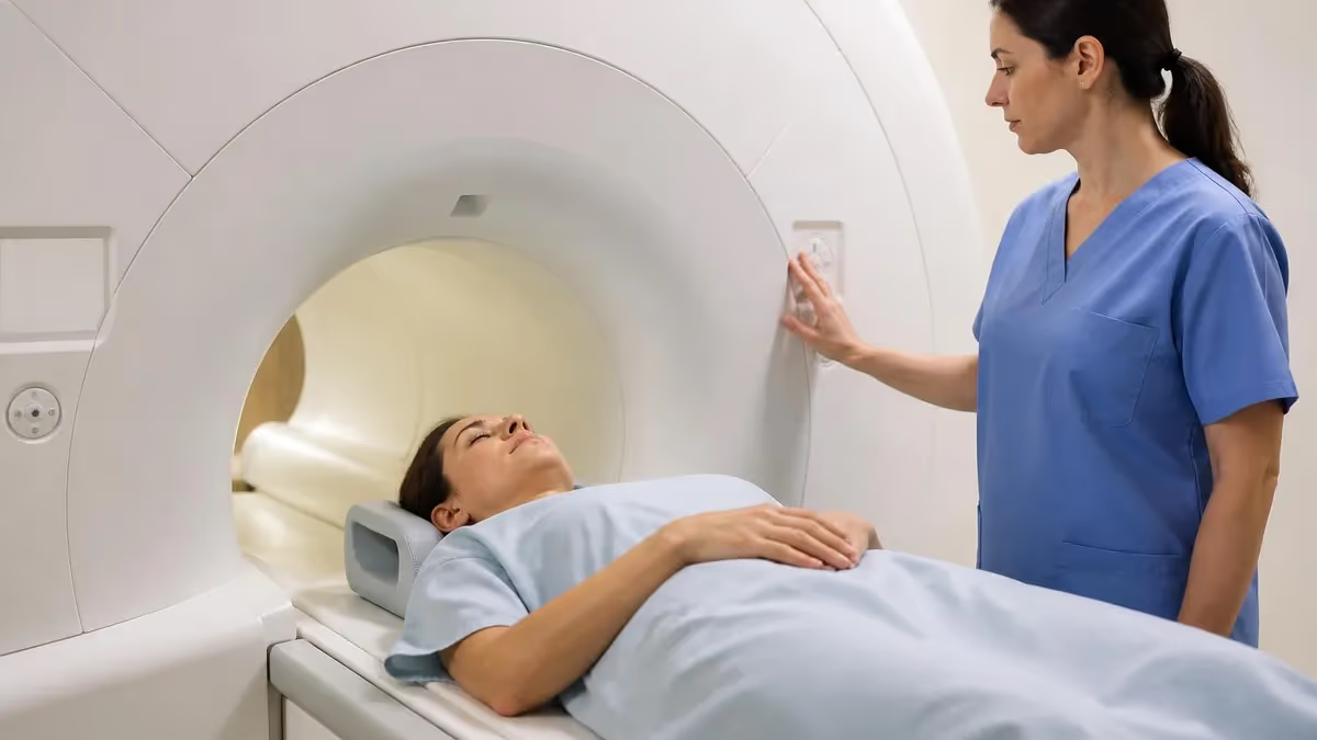
A liver MRI is the imaging study radiologists reach for when they need to know exactly what is going on inside the largest organ in your abdomen. Ultrasound is the first-line screening tool. CT is fast and widely available. But when the question gets harder - is that mass benign or malignant, how much iron has built up, how far has fibrosis progressed, why are the bile ducts dilated - MRI is the gold standard. It produces tissue contrast no other modality matches, and it does it without ionizing radiation.
If your doctor ordered a liver MRI, you are probably trying to make sense of why this scan and not something simpler. Maybe a routine ultrasound flagged a lesion that needs characterization. Maybe lab work showed elevated iron or unexplained liver enzymes. Maybe you are being staged for hepatocellular carcinoma surveillance because you have chronic hepatitis or cirrhosis. Each of those scenarios has its own protocol, contrast choices, and follow-up questions.
This guide walks through what a liver MRI actually shows, how the scan is built sequence by sequence, what hepatobiliary contrast agents do, how to prep, what the report jargon means, and how to read your bill. By the end you will know enough to walk into the imaging center with confident questions and walk out understanding what comes next.
Liver MRI by the Numbers
What does a liver MRI actually see? Think of the liver as a sponge of hepatocytes laced with blood vessels and bile ducts, all soaked in roughly the same kind of soft tissue. MRI exploits tiny differences in how protons relax in fat, water, iron-loaded cells, and abnormal tissue. Each sequence highlights a different property. Stack them together and you get a multi-dimensional read on what is normal, what is suspicious, and what is unambiguously pathologic.
The bread-and-butter findings on a liver MRI include hepatic steatosis (fatty liver), focal lesions like hemangiomas and adenomas, hepatocellular carcinoma (HCC) and metastatic deposits, iron overload from hemochromatosis or transfusion history, biliary obstruction, vascular abnormalities like portal vein thrombosis, and fibrosis grading using elastography sequences. A skilled radiologist can often distinguish a benign hemangioma from a malignant tumor on a single contrast-enhanced study, sparing the patient a biopsy that ultrasound or CT alone could not avoid.
One of the most useful aspects of liver MRI is dynamic post-contrast imaging. After gadolinium is injected, the scanner captures arterial, portal venous, and delayed phases within minutes. Each phase reveals different vessel and tissue behavior, and the pattern of enhancement and washout is often diagnostic by itself. Hepatocellular carcinoma classically lights up in the arterial phase and fades in the venous and delayed phases - a signature pattern called arterial enhancement with washout.
Key Insight
CT is faster, cheaper, and easier to schedule. But MRI delivers superior soft-tissue contrast, no ionizing radiation, and access to hepatobiliary contrast agents that specifically target liver cells. For patients who need repeat imaging - HCC surveillance, post-treatment follow-up, indeterminate lesion characterization - MRI's lack of radiation and superior tissue resolution earn it the lead role despite higher cost and longer scan time.
A typical liver MRI protocol stacks roughly six to ten sequences, each capturing a different tissue property. The technologist runs them back-to-back, sometimes with breath holds, sometimes with respiratory triggering for patients who cannot hold their breath. Total scan time usually lands between 30 and 60 minutes depending on whether contrast is used and which contrast agent your radiologist chose.
The opening sequences are non-contrast. T1-weighted in-phase and out-of-phase imaging detects microscopic fat - liver tissue that looks bright on in-phase but dims on out-of-phase has fatty infiltration even when ultrasound looks unremarkable. T2-weighted sequences highlight water content, making cysts and hemangiomas leap out as bright white. Diffusion-weighted imaging (DWI) flags malignant tissue by trapping water molecules; tumors restrict diffusion more than normal liver.
Then comes contrast. The radiologist will pre-specify either extracellular gadolinium (gadobutrol, gadoterate) or a hepatobiliary agent (gadoxetate, often sold as Eovist or Primovist). The latter is taken up by functioning hepatocytes about 20 minutes after injection, producing a hepatobiliary phase image where normal liver glows bright and non-hepatocyte lesions show as dark holes. This is the most sensitive sequence for detecting tiny metastases and characterizing focal nodular hyperplasia.
Core Sequences in a Liver MRI
Detects microscopic fat in hepatocytes by exploiting the chemical shift between water and fat protons. Drop in signal on out-of-phase imaging confirms steatosis.
Highlights water-rich structures. Cysts and hemangiomas appear very bright. Inflammation and edema also show up. Fat suppression keeps the surrounding tissue dark for contrast.
Measures how easily water molecules move through tissue. Tumors trap water and look bright on high b-value images, helping flag small malignant lesions.
Captures arterial, portal venous, and delayed phases after gadolinium injection. Patterns of enhancement and washout are often diagnostic of specific lesion types.
Liver MRI excels at characterizing focal lesions. A hemangioma - the most common benign liver tumor - shows peripheral nodular enhancement that fills in centripetally over time, a pattern so specific that biopsy is rarely needed. Focal nodular hyperplasia displays a central scar and strong arterial enhancement. Hepatic adenomas have a more variable look but often show signal drop on out-of-phase imaging because of intracellular fat. Each pattern tells a story the radiologist reads from a single multi-sequence study.
Hepatocellular carcinoma is where MRI truly shines as a screening and staging tool. The LI-RADS (Liver Imaging Reporting and Data System) algorithm categorizes lesions in cirrhotic livers from definitely benign to definitely malignant based on size, arterial enhancement, washout, capsule appearance, and threshold growth. A LI-RADS 5 lesion is diagnostic of HCC without biopsy - a remarkable example of imaging replacing tissue sampling. Patients on HCC surveillance for cirrhosis or chronic hepatitis B often get MRI every six months because the test is sensitive enough to catch tumors when they are still curable.
Diffuse liver disease shows up too. Iron overload from hemochromatosis or repeated transfusions makes the liver look dark on T2-weighted sequences. MR elastography measures tissue stiffness to grade fibrosis non-invasively, sometimes replacing biopsy entirely for patients with chronic hepatitis or non-alcoholic fatty liver disease. Bile duct disease - cholangiocarcinoma, primary sclerosing cholangitis, choledocholithiasis - is mapped beautifully by mri mrcp with and without contrast cpt code (MRCP), a fluid-sensitive sequence that draws bile ducts in white on a black background.
Common Reasons for a Liver MRI
Patients with cirrhosis or chronic hepatitis B carry elevated risk of hepatocellular carcinoma. Every six months, an ultrasound is the standard screen, but MRI is preferred when ultrasound is limited by body habitus, when AFP is rising without an ultrasound finding, or when a small nodule needs further characterization. LI-RADS scoring guides next steps.
The choice of contrast agent shapes the entire study. Extracellular gadolinium chelates - gadobutrol, gadoterate, gadobenate - distribute through blood vessels and interstitial spaces but do not get taken up by hepatocytes. They are excellent for vascular imaging, dynamic enhancement, and characterizing most lesions. Almost every imaging center stocks these because they are versatile and inexpensive.
Hepatobiliary agents are a special category. Gadoxetate disodium, marketed as Eovist in the United States and Primovist elsewhere, contains a gadolinium molecule attached to a transporter the liver actively pulls into hepatocytes. About 50 percent of the dose ends up in liver cells over the first 20 minutes. The remaining contrast circulates like a standard agent. After about 20 minutes, the scanner captures a hepatobiliary phase image where normal liver glows bright and any tissue not made of functioning hepatocytes - tumors, metastases, cysts, scar - shows up dark.
This hepatobiliary phase is the single most sensitive sequence for picking up small colorectal metastases, characterizing focal nodular hyperplasia (which contains hepatocytes and stays bright), and distinguishing HCC from regenerative nodules. The trade-off is longer scan time and higher cost, plus more frequent contrast-related transient dyspnea than with standard gadolinium. Your radiologist picks the agent based on the clinical question.
Gadolinium-based contrast agents have been linked to nephrogenic systemic fibrosis (NSF) in patients with severe kidney disease. Most imaging centers check estimated glomerular filtration rate (eGFR) before administering contrast. Patients with eGFR below 30 are usually screened out or given the lowest-risk agents (gadobutrol or gadoterate). Tell the technologist about any kidney disease, dialysis, or recent contrast exposure. Pregnancy and breastfeeding require separate discussion with your doctor.
Preparation for a liver MRI is more involved than for a brain or joint scan. Most protocols require four to six hours of fasting before arrival. Empty stomach reduces bowel motion artifact, decreases pancreatic and biliary secretion, and lets the gallbladder distend slightly so it can be evaluated. Some centers also have you drink a small amount of fluid contrast for the MRCP component, which makes the bowel signal disappear and improves bile duct visibility.
The screening form covers the usual MRI safety questions - implants, claustrophobia, pregnancy, prior contrast reactions, kidney function. For a liver scan, expect extra questions about recent abdominal surgery, biliary stents, drug pumps, transarterial chemoembolization beads, and any history of nephrogenic systemic fibrosis. The technologist places an IV line for contrast injection, usually in the antecubital vein, well before you enter the scanner room.
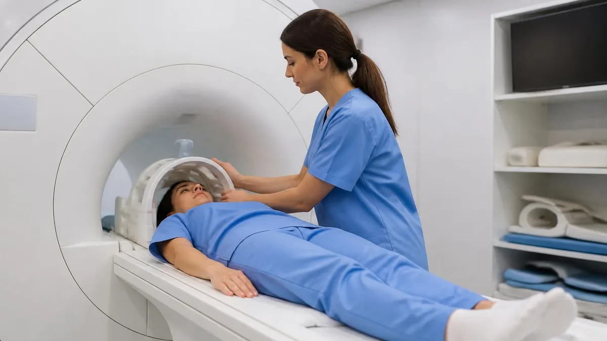
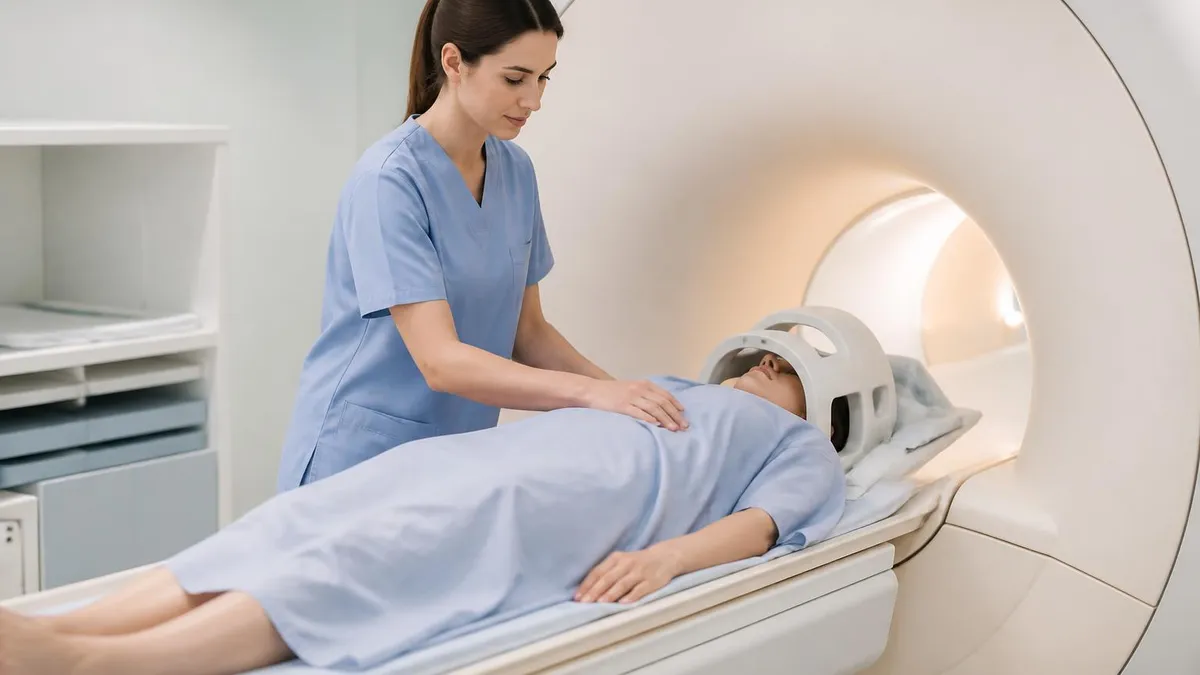
Inside the room, you lie supine on the table with a body coil wrapped around your torso. The technologist will coach you through breath holds - typically 15 to 20 seconds at a time. For patients who cannot hold their breath that long, respiratory-triggered sequences sync the scan to your breathing cycle, sacrificing some speed for tolerability. Headphones play music or instructions, and a squeeze ball lets you signal the tech if you need to stop.
Day-of-Scan Prep Checklist
- ✓Fast for 4 to 6 hours before the appointment (water in small sips usually allowed)
- ✓Bring photo ID, insurance card, and any prior imaging on disc
- ✓Have a recent eGFR or creatinine result available if contrast is planned
- ✓Wear loose clothes without metal zippers, snaps, or underwire
- ✓Remove jewelry, watches, hairpins, piercings, and dentures
- ✓List all surgeries, implants, and drug pumps with manufacturer details
- ✓Disclose any prior gadolinium reactions or contrast-induced shortness of breath
- ✓Practice 20-second breath holds at home if you have lung disease
- ✓Tell the tech if you are claustrophobic before entering the scanner room
- ✓Plan a ride home if mild sedation will be used for anxiety
Your radiology report will land in your patient portal within a day or two of the scan. Reading it can be disorienting because liver MRI uses precise but technical language. The structure is usually history, technique, findings, and impression. The impression is the bottom-line summary - if you read nothing else, read that. The findings section walks through each organ system observed on the scan and is where the radiologist documents both abnormalities and normal structures.
Common phrases worth understanding: a hyperintense or hypointense lesion describes signal brightness relative to surrounding liver on a specific sequence. Arterial hyperenhancement means a lesion lights up brighter than liver during the arterial phase of contrast. Washout describes a lesion that becomes darker than liver on portal venous or delayed phase imaging. A capsule appearance refers to a thin enhancing rim around a lesion. Persistent enhancement means the lesion stays bright into delayed phases, which can suggest hemangioma or other benign findings.
If LI-RADS scoring is used, expect categories from LR-1 (definitely benign) through LR-5 (definitely HCC), plus LR-M (probably or definitely malignant but not necessarily HCC) and LR-TIV (tumor in vein). Each category triggers different follow-up - imaging surveillance for low scores, biopsy or treatment for high scores. The system is standardized so reports from different institutions communicate the same risk levels.
Bring questions to your follow-up appointment with the ordering physician. Plain Google searches on radiology terms can spiral into worst-case scenarios that have nothing to do with your actual situation. Your doctor combines the report with labs, history, physical exam, and prior imaging to arrive at the real interpretation. A finding that sounds alarming in isolation often means very little once context is added.
Liver MRI Pros and Cons
- +No ionizing radiation - safe for repeated surveillance scans
- +Superior soft-tissue contrast over CT and ultrasound
- +Hepatobiliary contrast agents specifically map liver function
- +MR elastography measures fibrosis non-invasively
- +MRCP draws bile ducts without invasive procedures
- +Can characterize most lesions without biopsy
- −Longer scan time, often 45 minutes or more
- −Higher cost than CT or ultrasound
- −Requires breath holds that can be hard for some patients
- −Not suitable for patients with certain implants
- −Gadolinium contraindicated in severe kidney disease
- −Image quality degrades with patient motion or ascites
How much does a liver MRI cost? In the United States, prices vary wildly. A freestanding imaging center may quote $500 to $1,200 cash for a non-contrast study, $800 to $2,000 with extracellular gadolinium, and $1,500 to $3,000 if hepatobiliary contrast like Eovist is used. Hospital outpatient departments often charge two to three times those numbers for the same scan on similar equipment. The CPT codes (74181, 74182, 74183) reflect with-or-without contrast distinctions but do not differentiate hepatobiliary from extracellular agents, which is why the latter often appears as a separate line-item charge.
Insurance coverage is generally available when the scan is medically necessary, but most plans require prior authorization. The ordering physician's office submits clinical justification - elevated AFP, indeterminate ultrasound finding, cirrhosis surveillance schedule, oncologic staging - and the insurer either approves, denies, or asks for a peer-to-peer review. This process can delay scheduling by several days to several weeks. If your case is urgent, ask the ordering office to flag the request and expedite.
If you are paying cash or have a high-deductible plan, shop aggressively. Get the CPT code from the order, call three or four imaging centers within driving range, and ask for the cash-pay rate. Some centers post prices online. Hospital outpatient departments are almost always more expensive than freestanding centers running identical equipment. The radiologist reading the scan does not have to be at the imaging center - many studies are interpreted by subspecialty teleradiology groups, so location matters less than you might think.
For HCC surveillance specifically, ask your hepatologist whether MRI or ultrasound is appropriate every six months. Some patients qualify for MRI surveillance based on body habitus, ultrasound limitations, or insurer policy. Others stay on ultrasound and only escalate to MRI when something looks suspicious. The right strategy is patient-specific and worth a clear conversation up front rather than discovering coverage gaps after a scan.
MRI Questions and Answers
A liver MRI gives radiologists a multi-dimensional view of the largest organ in your abdomen, combining structural detail, tissue characterization, and functional information no other single test can match. For HCC surveillance, indeterminate lesion characterization, pre-surgical planning, and fibrosis assessment, it is the most powerful tool in the imaging arsenal. The trade-off - longer scan times, higher cost, more involved prep - is usually worth it for the diagnostic certainty the study delivers.
If you are preparing for your own liver MRI, the practical takeaways are simple. Fast as instructed. Disclose kidney problems and prior reactions. Practice breath holds if you have lung disease. Plan for an hour in the scanner. Ask your doctor whether hepatobiliary contrast adds enough value to justify the higher cost in your case. Request your images and report afterward and bring them to follow-up visits as prior comparisons for any future studies.
If you are studying for the MRI registry, liver protocols are a high-yield exam topic. Know the in-phase versus out-of-phase signal behavior, the dynamic post-contrast phases, the role of MRCP, and the difference between extracellular and hepatobiliary contrast. LI-RADS basics show up on board questions, as do the safety considerations around gadolinium and renal function. Pair concept review with hundreds of practice questions and the patterns will stick.
Imaging keeps advancing. Abbreviated MRI protocols are shrinking HCC surveillance scans to 10 or 15 minutes by skipping non-essential sequences. Deep-learning reconstruction is denoising images so scans finish faster without losing detail. Photon-counting CT is starting to challenge MRI for some indications. But for the foreseeable future, when a hepatologist needs to know what is happening inside a liver with the highest possible confidence and the lowest possible risk, the answer is a multi-sequence multi-phase MRI - and you now know exactly what that means and what to expect.
About the Author

Medical Laboratory Scientist & Clinical Certification Expert
Johns Hopkins UniversityDr. Sandra Kim holds a PhD in Clinical Laboratory Science from Johns Hopkins University and is certified as a Medical Technologist (MT) and Medical Laboratory Scientist (MLS) through ASCP. With 16 years of clinical laboratory experience spanning hematology, microbiology, and molecular diagnostics, she prepares candidates for ASCP board exams, MLT, MLS, and specialist certification tests.
Join the Discussion
Connect with other students preparing for this exam. Share tips, ask questions, and get advice from people who have been there.
View discussion (8 replies)