Heart MRI: What It Shows, How It Works, and What to Expect 2026 July
Heart MRI guide: ⏳ what cardiac MRI shows, prep, scan steps, contrast safety, results, costs vs echo & CT, and how long it takes.
A heart MRI uses a powerful magnetic field and radio waves to map the chambers, valves, and muscle of your heart in remarkable detail. Cardiologists often order this scan when an echocardiogram leaves questions unanswered or when a structural problem needs sharper imaging. Unlike a CT scan, a heart MRI uses no ionizing radiation, which matters if you need follow-up scans across many years.
The exam is also called cardiac magnetic resonance, or CMR for short. Radiologists use it to study how blood flows through each chamber, how thick the heart muscle has become, and whether scar tissue is hiding in places a standard ultrasound cannot reach. The pictures show motion, structure, and tissue character at the same time, which is why this single test can replace several older ones.
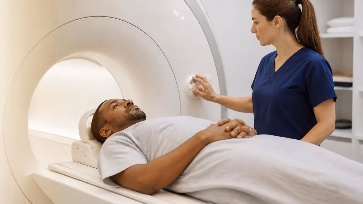
If your doctor mentioned a heart MRI, you probably want to know what to expect. The scan takes longer than a knee or shoulder study and asks you to hold your breath at points. None of it hurts. The magnet is strong, but the field itself is invisible and silent until the gradient coils start their loud, rhythmic knocking.
What a Heart MRI Actually Shows
A heart MRI captures the heart in motion. Cine sequences acquire dozens of frames across the cardiac cycle and then stitch them together so the radiologist can watch your heart pump in real-time playback. From those clips the team measures ejection fraction, chamber volumes, and wall thickness with accuracy that echocardiography struggles to match.
The scan also exposes scar tissue. After a gadolinium contrast injection, healthy muscle washes the agent out quickly while damaged tissue holds onto it. That delayed enhancement pattern reveals old heart attacks, inflammation from myocarditis, and the streaky fibrosis seen in hypertrophic cardiomyopathy. Many of these findings stay invisible on every other imaging test.
Valve function shows up too. Phase-contrast sequences measure blood velocity through the aortic and mitral valves, so the radiologist can quantify leaks and narrowings in milliliters per beat. The result reads more like a flow chart than a fuzzy ultrasound clip, which is why surgical teams lean on cardiac MRI before valve repair or replacement.
Heart MRI by the Numbers
Why Your Doctor Ordered the Scan
There are a handful of common reasons. The first is suspected cardiomyopathy, where the heart muscle has thickened, dilated, or stiffened without an obvious cause. A CMR can sort hypertrophic from infiltrative disease in a single appointment, which changes the entire treatment plan. The second is after a heart attack, when surgeons need to know how much muscle is salvageable before considering a bypass.
Other patients arrive with arrhythmias of unknown origin. The MRI can find tiny patches of scar that trigger ventricular tachycardia and which ablation might target. Athletes get scanned when an ECG shows abnormal voltages but the echocardiogram looks clean. A small subset receive cardiac MRI for congenital heart disease follow-up, especially adults who had surgery as infants and now need an updated map of their anatomy.
If you have implanted devices, the conversation gets longer. Most modern pacemakers and defibrillators are now MRI conditional, but the imaging team has to confirm the model, reprogram it, and monitor your rhythm during the scan. Read the full breakdown in our guide to MRI with pacemaker before your appointment.

Key Point: No Radiation
Unlike a coronary CT or nuclear stress test, a heart MRI uses no ionizing radiation. This matters for younger patients, women in pre-menopause, and anyone who needs repeat imaging across many years.
Before You Arrive: Prep Steps That Matter
Most patients can eat and drink as usual. The exception is when stress-perfusion imaging is on the schedule, which is a pharmacologic stress test done inside the magnet. For that protocol the technologist will ask you to avoid caffeine for 24 hours because coffee, tea, chocolate, and many sodas block the medication used to dilate your coronary arteries.
Bring a list of every implant, surgical clip, or piece of retained metal. Some old aneurysm clips and cochlear implants are absolute contraindications. Your screening form will ask twice, and the technologist will ask again at the magnet door. This is not paranoia. The magnet stays on, all the time, and ferromagnetic objects become projectiles.
Wear something with no metal. Many imaging centers issue scrubs to remove any doubt. Leave watches, jewelry, and credit cards in the locker. Hearing aids stay outside the scanner room. So do CPAP masks unless the team confirms a model that is safe for the bore.
Heart MRI Prep Checklist
- ✓Skip caffeine for the full 24 hours before your scan if it includes stress perfusion imaging since caffeine blocks the medication used to dilate coronary arteries
- ✓Wear clothing with no metal zippers, snaps, or wires, or plan to change into scrubs at the imaging center for safety reasons
- ✓Bring a complete list of every implant, past surgery, and any retained metal fragments from prior injuries or industrial work
- ✓Bring the device card for any pacemaker, defibrillator, neurostimulator, or insulin pump so the team can confirm MRI conditional status
- ✓Stay well hydrated the day before and the day after the scan to help your kidneys clear the gadolinium contrast efficiently
- ✓Plan for 45 to 90 minutes on the table, plus an additional hour for prep work like IV placement and ECG lead attachment
- ✓Arrange a quiet room afterward if you took a sedative for claustrophobia and bring someone to drive you home
- ✓Take all regular medications unless your cardiologist specifically tells you to hold a dose for the day of the scan
Inside the Scanner: What Happens Step by Step
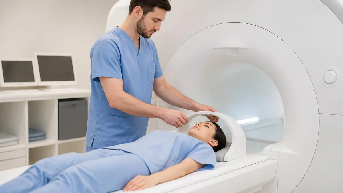
You lie on your back on a padded table. The technologist places a flexible coil over your chest. This coil receives the radio signal from your heart and feeds it back to the computer. Most patients also get adhesive electrodes for an MRI-compatible ECG, which lets the computer time each image to the same point in your cardiac cycle.
The table slides into the bore. The opening is roughly 60 to 70 centimeters wide depending on the magnet, and your head and shoulders sit inside it. Wide-bore systems feel less confining than older closed magnets. If claustrophobia worries you, ask about the wide bore MRI option at your imaging center.
The technologist talks to you through an intercom and runs short sequences first to plan the rest of the study. Then come the cine acquisitions. You will hear instructions to breathe in, breathe out, and hold for about 10 to 15 seconds. Each breath-hold captures one slice or one stack of frames. The knocking can climb above 100 decibels during fast sequences, which is why you receive earplugs or headphones.
Roughly halfway through, the nurse or technologist injects gadolinium contrast through a small cannula in your arm. The agent flushes through your heart in about 60 seconds and improves the contrast between blood, muscle, and scar. Some people taste metal briefly. A cool sensation travels up the arm. Neither lasts more than a minute.
Three Things a Heart MRI Measures Better Than Any Other Test
Cine sequences acquire dozens of frames per heartbeat. The software then traces the inner border of each chamber to calculate volume and ejection fraction with high accuracy.
After gadolinium injection, scar tissue holds onto the contrast while healthy muscle clears it. The bright streaks on late enhancement images reveal old heart attacks and fibrosis.
Phase-contrast sequences measure blood velocity through the valves. The radiologist can quantify leaks and narrowings in milliliters per beat, not just visual estimates.
How Long a Heart MRI Takes
Plan for 45 to 90 minutes on the table. A focused viability study runs shorter. A full work-up with cine, perfusion, late gadolinium enhancement, and flow imaging fills the upper end of that window. If you want comparisons with other regions, our guide on how long does an MRI take breaks down scan times by body part.
Add another 30 minutes for prep. The cannula has to be placed, the ECG leads attached, and the consent forms reviewed. Stress protocols add another 15 to 20 minutes because the medication has to clear before the late images. Most centers ask you to arrive an hour before the scheduled start.
Gadolinium Contrast: Is It Safe?
Gadolinium is the contrast agent used in nearly every cardiac MRI. It shortens the relaxation time of nearby tissue, which is how scar tissue lights up on late enhancement images. Modern macrocyclic agents are stable, clear through the kidneys, and have an excellent safety record across millions of doses.
The main caution is for people with significant kidney impairment. Patients with an estimated GFR below 30 mL/min face a small but real risk of nephrogenic systemic fibrosis with older linear agents. Macrocyclic agents have nearly eliminated that risk, but most departments still check your renal function before injecting.
Allergic reactions are rare. Mild flushing or a brief headache happens to maybe 1 in 100 patients. Severe reactions are vanishingly uncommon, in the range of 1 in 10,000. The contrast room keeps emergency medications on hand for the rare event. Pregnancy is the other limit, since gadolinium crosses the placenta. Read more on MRI and pregnancy if this applies to you.
Pacemakers, defibrillators, cochlear implants, aneurysm clips, insulin pumps, and some dental hardware all matter. Some are absolute contraindications. Some are MRI conditional with specific safety steps. Bring the device card if you have one.
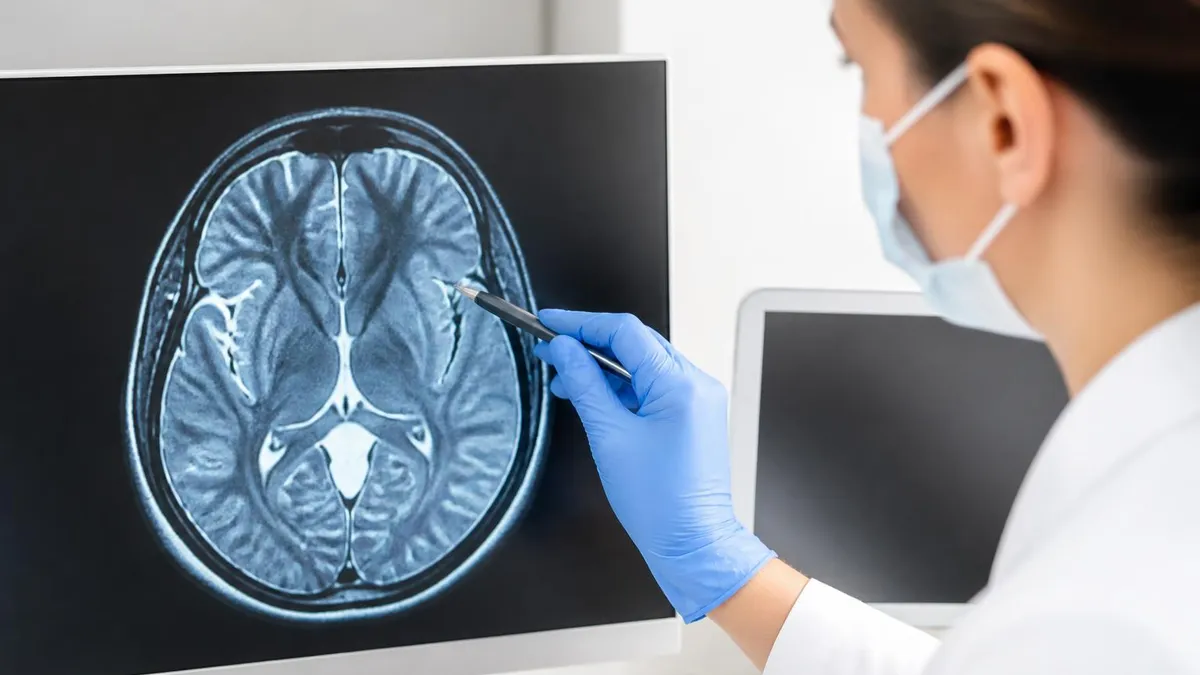
What the Results Tell Your Cardiologist
The radiologist sends a structured report to your cardiologist within a day or two at most centers. It covers chamber sizes, ejection fraction of both ventricles, valve function in numerical terms, and any pattern of enhancement. The cardiologist then matches those findings to your symptoms, your ECG, and your blood work.
Ejection fraction is the headline number for many patients. A normal left ventricle pumps out 55% or more of its blood with each beat. Values in the 40% range suggest mild dysfunction. Below 35% you enter the territory where defibrillators and certain medications get serious consideration. The CMR number is more accurate than the echocardiogram value and often changes treatment.
Late enhancement patterns guide diagnosis. Subendocardial enhancement that follows a coronary artery distribution points to an old heart attack. Mid-wall striae suggest non-ischemic cardiomyopathy. Patchy mid-wall and subepicardial enhancement raises the possibility of myocarditis. Each pattern carries a different treatment path.
What Different MRI Sequences Show
Steady-state free precession captures the heart in motion across the cardiac cycle. These are the bright, high-contrast clips that look like a real beating heart on playback. They are used for chamber size, wall motion, and ejection fraction. The radiologist draws inner contours around each chamber on end-diastolic and end-systolic frames to calculate volumes.
Risks, Limits, and Honest Trade-offs
The MRI itself is non-invasive and uses no radiation. The main risks come from the contrast and from missed metal. Both are managed by screening. The bigger limit for many patients is the breath-holding requirement. People with significant lung disease, severe heart failure, or persistent arrhythmias may not be able to hold long enough to get clean cine images. In that case the team may switch to a free-breathing protocol that uses navigator gating, but image quality drops.
Cost is another factor. A heart MRI is more expensive than an echocardiogram and varies widely by region and facility. Our guide on MRI scan cost breaks down typical prices with and without insurance. If you have a high deductible plan, ask for the cash price up front. Hospital outpatient facilities often charge two to three times what a freestanding imaging center bills.
Image quality also depends on the magnet. A 1.5 Tesla system handles most cardiac work well. A 3 Tesla system gives a stronger signal but is more sensitive to arrhythmia artifact. The choice matters less for routine viability studies and more for delicate flow quantification.
Heart MRI vs Echocardiogram vs CT Angiography
Each test answers a different question. Echocardiography is fast, cheap, portable, and uses ultrasound waves with no radiation. It is the right first test for most patients. Coronary CT angiography uses X-rays and iodinated contrast to map the coronary arteries themselves. Heart MRI offers neither speed nor coronary detail, but it dominates in three areas: chamber quantification, tissue characterization, and complex flow.
If your cardiologist needs to know whether your heart muscle is scarred, inflamed, or infiltrated by amyloid, MRI is the test. If the question is whether your coronary arteries are blocked, CT angiography usually wins. If the question is simply about ejection fraction and valve flow, an echo may be enough. Many patients end up with two or three of these tests over a year of work-up.
After the Scan: What Comes Next
You can drive yourself home unless you received a sedative for claustrophobia. Drinking extra water helps your kidneys clear the gadolinium over the next 24 hours. There are no activity restrictions. The cannula puncture site is usually fine within an hour.
Your cardiologist usually schedules a follow-up visit within a week or two to review the findings. Bring questions. Ask what the ejection fraction was, whether any enhancement was seen, and what the next step is.
For some patients the heart MRI is the end of the workup. For others it is the start of a longer plan that includes medication adjustments, an implantable device, or surgery. Either way, you walked out with one of the most detailed maps of your heart that medicine can produce, and that map will guide decisions for years.
Common Reasons a Heart MRI Is Ordered
When the heart muscle has thickened, dilated, or stiffened without an obvious cause, CMR sorts hypertrophic from infiltrative disease in one appointment.
Surgeons need to know how much muscle is salvageable before considering bypass or angioplasty. Late enhancement maps the dead tissue precisely.
MRI can find tiny scar patches that trigger ventricular tachycardia. Those targets help guide ablation procedures.
Adults who had surgery as infants need an updated map of their anatomy. CMR is the imaging tool of choice for these patients.
Special Situations You Should Know About
Athletes occupy a special category in cardiac imaging. The athletic heart adapts to training by enlarging and increasing wall thickness, and those changes can look concerning on an echocardiogram. A heart MRI sorts out true cardiomyopathy from physiologic adaptation by looking at the pattern of enhancement and the symmetry of wall thickness. A clean late enhancement scan in a thickened heart usually points to training rather than disease.
Pregnancy raises different questions. The magnet itself is considered safe across all trimesters, although elective scans usually wait until after delivery. Gadolinium contrast is generally avoided because it crosses the placenta. If a heart MRI is medically necessary during pregnancy, most centers run a non-contrast protocol. Your obstetrician and cardiologist will coordinate the decision together.
Children get cardiac MRI too, especially when they were born with structural heart disease and need follow-up imaging as they grow. Pediatric protocols are shorter, the breath-holding requirement is replaced with free-breathing techniques in younger kids, and sedation may be used for toddlers who cannot stay still.
Common Findings Explained in Plain Language
Reading a cardiac MRI report can feel like decoding a foreign language. Here are the terms that show up most often. Wall motion abnormality means a segment of the heart muscle is not contracting normally. This often correlates with an old heart attack or with active ischemia. The report will name the wall and segment, like inferior, septal, or anterolateral.
LV mass index is the weight of the left ventricle, adjusted for body size. Elevated values appear in hypertension, in athletes, and in genetic conditions. The report compares your number to a reference range and may note whether the thickening is concentric or asymmetric. Asymmetric septal thickening is the hallmark of hypertrophic cardiomyopathy.
Right ventricular function gets its own paragraph. The right ventricle is harder to image with ultrasound, which is one of the major advantages of CMR. A reduced right ventricular ejection fraction shows up in pulmonary hypertension, in arrhythmogenic right ventricular cardiomyopathy, and in late stages of left-sided heart failure.
MRI Questions and Answers
About the Author

Medical Laboratory Scientist & Clinical Certification Expert
Johns Hopkins UniversityDr. Sandra Kim holds a PhD in Clinical Laboratory Science from Johns Hopkins University and is certified as a Medical Technologist (MT) and Medical Laboratory Scientist (MLS) through ASCP. With 16 years of clinical laboratory experience spanning hematology, microbiology, and molecular diagnostics, she prepares candidates for ASCP board exams, MLT, MLS, and specialist certification tests.
Join the Discussion
Connect with other students preparing for this exam. Share tips, ask questions, and get advice from people who have been there.
View discussion (6 replies)