MRI Principles: The Physics and Imaging Science Behind the Scan
MRI principles explained: nuclear magnetic resonance, T1 and T2 relaxation, gradients, RF pulses, and how MRI builds images of the body.
At a Glance: MRI principles rest on nuclear magnetic resonance. A static magnetic field aligns hydrogen protons, a radio frequency pulse tips them sideways, and three gradient coils encode position. The relaxation signal is then transformed by Fourier mathematics into diagnostic cross-sectional images, all without ionizing radiation.
MRI principles begin with one fact. Most hydrogen in your body sits inside water and fat. Place that hydrogen inside a strong magnetic field and the tiny nuclear spins line up. Send a tuned radio wave at them and they tip sideways, then relax back. That relaxation signal is what the scanner listens for. It is the foundation of every protocol used on a 3T MRI system today.
The technical name is nuclear magnetic resonance. Protons in a magnetic field precess at the Larmor frequency. At 1.5 tesla that frequency is about 63.8 MHz. At 3 tesla it doubles to roughly 127.7 MHz. The scanner broadcasts radio pulses tuned to that exact frequency. MRI uses radio waves rather than X-rays, so no ionizing radiation is delivered to the body. That is why a baby MRI is preferred for sensitive populations whenever clinical questions can be answered without contrast.
Each MRI image is built from three magnetic gradients layered on top of the main field. The slice-selection gradient picks the imaging plane. The phase-encoding gradient labels each row of voxels with a unique phase shift. The frequency-encoding gradient labels each column with a unique frequency. A mathematical Fourier transform then sorts these phases and frequencies into a grid of pixels. The same principle works for a cervical MRI or a focused ankle study.
MRI by the Numbers

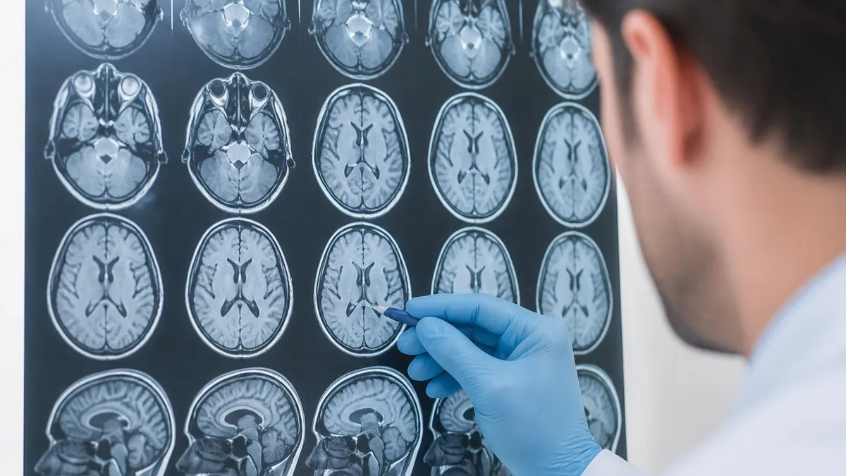
Contrast in MRI comes from two relaxation times called T1 and T2. T1 measures how fast spins recover along the main magnetic field after a pulse. T2 measures how fast spins lose phase coherence in the transverse plane. Fat has a short T1 and looks bright on T1-weighted scans. Water has a long T1 and T2. Radiologists use the differences to spot tumors, edema, and hemorrhage on a brain MRI or spine study.
Beyond T1 and T2, MRI offers a toolbox of weighting options. Proton density imaging emphasizes how many hydrogen atoms sit in each voxel. FLAIR suppresses cerebrospinal fluid so lesions near the ventricles stand out from background. STIR suppresses fat so bone marrow edema jumps out on musculoskeletal exams. Diffusion-weighted imaging maps how freely water molecules move, which is how a stroke appears within minutes of onset.
The Three Gradient Coils
Picks the imaging plane by varying field strength along one axis. Only protons at the chosen position resonate with the RF pulse, defining slice thickness and location.
Applied briefly between excitation and readout. Labels each row of the matrix with a unique phase shift, which is decoded by the Fourier transform during reconstruction.
Switched on during signal readout. Causes protons in each column to precess at a slightly different frequency, providing spatial mapping along the readout direction.
Gadolinium contrast agents change the local magnetic environment to shorten T1. Areas that collect the contrast, like inflammation or tumor neovascularity, become bright on T1 after injection. That is why a brain MRI with contrast is more sensitive for multiple sclerosis plaques and small metastases than a non-contrast study. Contrast is delivered through a small IV at roughly 0.1 mmol per kilogram of body weight. It is renally cleared within about 24 hours in healthy patients with normal renal function.
Some clinical settings call for hepatobiliary contrast such as gadoxetate disodium, which is taken up by functioning hepatocytes. This lets radiologists distinguish focal nodular hyperplasia from adenomas during a liver MRI. Other studies use ferumoxytol, an iron-based agent originally approved for anemia, as a blood-pool MR contrast. Each agent works because of the same underlying physics that gives every MRI exam its diagnostic power.
Common MRI Sequences
Short TR and short TE. Fat appears bright, water appears dark. Best for anatomy and post-contrast imaging. Shows subacute hemorrhage as bright signal. The workhorse anatomic sequence in nearly every brain, spine, and joint protocol.
Spatial resolution depends on matrix size, field of view, and slice thickness. A typical knee study uses a 320 by 256 matrix over a 16 cm field of view with 3 mm slices. That yields voxels around 0.5 by 0.6 by 3 mm. Smaller voxels capture finer anatomy but lower the signal-to-noise ratio, which means longer scan times. The same trade-off applies to a knee MRI, a wrist study, or a high-resolution pituitary protocol.
Signal-to-noise ratio, or SNR, sits at the heart of every MRI decision. SNR rises with field strength, with the number of signal averages, and with the size of the receive coil elements. A 3T system roughly doubles SNR compared to 1.5T at the same scan parameters. That is why ultra-fine neuro and musculoskeletal exams often happen on 3T magnets. Dedicated coils for spine, breast, and small joints pull more signal from the body and shorten scans.
Patients with severe kidney impairment, with an eGFR below 30 mL/min/1.73 m2, face a higher risk of nephrogenic systemic fibrosis. Always check renal function before injecting gadolinium contrast and follow your institution's protocol for screening and consent.
Echo planar imaging is one of the most important fast-scan techniques in modern MRI. Instead of acquiring one line of k-space per excitation, EPI fills the whole matrix after a single radio pulse. It uses rapidly switching gradients to do so. This makes diffusion-weighted imaging, perfusion imaging, and functional MRI possible in seconds. The cost is sensitivity to magnetic field inhomogeneity near sinuses and metal hardware.
Parallel imaging accelerates scans by using spatial information already encoded in multi-channel receive coils. Methods such as SENSE, GRAPPA, and CAIPIRINHA undersample k-space and then reconstruct missing data from coil sensitivity maps. A factor-of-two acceleration cuts scan time in half. That speed gain is essential for breath-held abdominal imaging, cine sequences, and dynamic contrast studies on a modern cardiac MRI exam. Higher accelerations are possible with newer 32 and 64-channel coil designs.
MRI Safety Screening Checklist
- ✓Pacemaker, defibrillator, or implanted cardiac device check
- ✓Cerebral aneurysm clip or carotid stent documentation
- ✓Cochlear implant or middle-ear prosthesis history
- ✓Metal fragments in the eye from welding, grinding, or trauma
- ✓Insulin pump, neurostimulator, or drug-infusion device
- ✓Pregnancy status, particularly first trimester exposure
- ✓Tattoos with metallic pigments or recent permanent makeup
- ✓Renal function adequate for gadolinium contrast administration
Safety is built into every MRI principle. The static magnetic field never turns off. Ferromagnetic objects accelerate toward the bore at lethal speed. That is why screening forms ask about pacemakers, aneurysm clips, cochlear implants, and shrapnel. The gradient coils generate the loud knocking sound and can stimulate peripheral nerves at high amplitudes. The radio frequency pulses deposit heat in tissue, measured as the specific absorption rate, or SAR.
Artifacts tell the technologist that a physics principle is being violated somewhere in the imaging chain. Wraparound, or aliasing, happens when the field of view is smaller than the body part. Chemical shift artifact appears at fat-water interfaces because fat and water precess at slightly different frequencies. Motion artifact streaks across the phase-encoding direction because that axis is sampled over many seconds. Recognizing these patterns is part of being confident on a full body MRI or focused brain study.
Image reconstruction is no longer a simple Fourier transform. Compressed sensing exploits the fact that MRI images are sparse in some mathematical domain. Iterative algorithms then recover the image from heavily undersampled data. Deep-learning reconstructions trained on large datasets now denoise and sharpen scans in real time. These advances let modern cervical spine MRI studies finish in half the time of a decade ago.
K-space is the workhorse abstraction every MRI physicist uses to describe data acquisition. Each row corresponds to a different phase-encoding step. Each column corresponds to a different frequency sample within that step. Low-frequency points in the center of k-space encode contrast. High-frequency points around the edges encode fine detail. Acquiring the center first gives stronger contrast in motion-corrupted scans. Skipping outer lines speeds the exam but blurs sharp edges of anatomy.
1.5T vs 3T MRI Systems
- +Higher SNR for fine anatomy and small structures
- +Faster scans at equal resolution and matrix
- +Stronger functional and spectroscopy signal
- +Sharper diffusion and perfusion imaging quality
- −Stronger chemical shift artifact at tissue interfaces
- −Greater susceptibility distortion near metal implants
- −Higher SAR limits scan parameters and TR options
- −Higher cost per minute of scanner time and maintenance
Echo time, repetition time, and flip angle are the three knobs every tech turns at the console. Echo time, or TE, sets when the signal is sampled after the excitation pulse and controls T2 weighting. Repetition time, or TR, sets how often the sequence repeats and controls T1 weighting. Flip angle controls how far the spins are tipped, balancing SNR against scan time. Memorize the rule that short TR plus short TE equals T1 weighting on any lumbar MRI protocol.
Coil selection separates good images from great ones. Surface coils sit close to the anatomy and capture strong signal but only over a limited depth. Volume coils such as the body coil image larger regions but with less SNR per voxel. Modern scanners combine multiple small elements into a phased array, giving both wide coverage and high SNR. For a breast MRI, dedicated breast coils with 8 to 32 channels enable bilateral high-resolution imaging in one acquisition.
Fast-Scan Techniques
Fills k-space after a single RF pulse using rapid gradient switching. Powers diffusion, perfusion, and functional MRI in seconds. Sensitive to field inhomogeneity near sinuses and metal.
Undersamples k-space and reconstructs missing data using coil sensitivity maps. SENSE, GRAPPA, and CAIPIRINHA cut scan time by a factor of two or more without major SNR loss.
Exploits the natural sparsity of MR images in transform domains. Allows acquisition of only a fraction of k-space lines and iterative reconstruction recovers full-resolution images.
Neural networks trained on large image datasets denoise and sharpen scans during reconstruction. Modern scanners ship with vendor implementations for routine clinical use.
Magnetic field homogeneity, called shimming, is tweaked before every exam. Imperfect homogeneity creates dark bands across the image, breaks fat suppression, and distorts diffusion scans. Active shimming uses extra coils carrying small currents to flatten the main field. Passive shimming uses iron plates placed inside the bore at installation. Higher field strength magnets such as 3T systems require more careful shimming, particularly around the skull base and abdomen.
Functional MRI takes advantage of blood oxygenation level dependent contrast. When neurons fire, local blood vessels deliver more oxygenated blood than the tissue actually consumes. Oxygenated hemoglobin is diamagnetic. Deoxygenated hemoglobin is paramagnetic. The net result is a small increase in T2-star signal in active brain regions. Repeated EPI volumes acquired during tasks map language, motor, and memory networks for neurosurgical planning at major centers.
MR angiography images blood vessels without injecting iodinated contrast. Time-of-flight angiography relies on the bright signal of fresh, unsaturated blood flowing into a slice. Phase-contrast angiography uses bipolar gradients to encode flow velocity in the phase of the signal. Contrast-enhanced MRA injects a bolus of gadolinium and times acquisition to peak arterial enhancement. Each technique tells a different story about anatomy, flow direction, and velocity.
Typical Sequence Parameters
Quality control rounds out the principles you need to understand. Daily phantom scans verify SNR, geometric accuracy, slice thickness, and ghosting levels. Weekly checks confirm gradient calibration is on target. Quarterly tests measure coil performance and image uniformity across the bore. Annual surveys by medical physicists document drift in field strength and shim quality on every clinical scanner.
Understanding MRI principles helps you pass certification exams and feel confident at the console. Practice that knowledge with realistic question banks where each item explains the physics behind the right answer. Pair that with hands-on time scanning real patients. You will start to see how every protocol choice, from echo time to receiver bandwidth, traces back to a small set of physical laws. That mastery is the difference between simply running a scanner and truly understanding it.
Cross-talk between adjacent slices is a subtle physics issue every tech should understand. A real RF pulse has a slightly imperfect slice profile that bleeds energy into neighboring slices. If those slices are imaged in immediate sequence, their longitudinal magnetization has not fully recovered. That lowers signal and changes contrast in unwanted ways. Interleaving slice order, leaving small gaps between slices, or using a 3D acquisition that excites a whole slab at once are the three standard ways to manage cross-talk in any clinical protocol.
Three-dimensional sequences acquire a volume of tissue in one go rather than slice by slice. Examples include MPRAGE for brain anatomy, VIBE for the abdomen, and CISS for the inner ear and cranial nerves. Each 3D scan delivers thin contiguous slices that can be reformatted in any plane without losing resolution. The trade-off is longer scan times and tighter motion sensitivity. That is why 3D scans usually require breath-holding or motion-correction techniques to produce a diagnostic MRI study.
Susceptibility-weighted imaging exploits the magnetic differences between materials such as deoxygenated blood, iron, calcium, and air. These materials distort the local magnetic field and create phase shifts that are amplified in a special post-processing step. The result is exquisite sensitivity to microbleeds, cerebral cavernous malformations, and small calcifications that other sequences miss. Susceptibility-weighted imaging has become a routine part of trauma, stroke, and dementia protocols at most centers. It adds critical information without lengthening the exam by more than a few minutes per study.
Magnetization transfer is one more contrast mechanism worth understanding. Protons bound to large macromolecules have very short T2 and do not show up in standard images. Applying an off-resonance pulse saturates these bound protons. The saturation transfers to the free water pool through chemical exchange and dipole coupling, reducing its signal. Tissues rich in macromolecules, such as white matter and cartilage, lose signal more than tissues such as cerebrospinal fluid, which highlights demyelination in multiple sclerosis monitoring.
Advanced MRI Applications
Measures concentrations of brain metabolites such as N-acetylaspartate, choline, creatine, and lactate. Used to grade tumors and distinguish recurrence from radiation necrosis without biopsy.
Exploits magnetic differences between deoxyhemoglobin, iron, calcium, and air. Highly sensitive to microbleeds, cavernous malformations, and small calcifications in trauma and stroke.
Off-resonance pulses saturate macromolecule-bound protons. Loss of signal in white matter and cartilage helps quantify demyelination in multiple sclerosis monitoring.
Measures water diffusion along multiple directions. Tractography reconstructions reveal white matter tracts for neurosurgical planning and study of brain connectivity.
Spectroscopy is a niche but important application that uses the same nuclear magnetic resonance principles to measure metabolite concentrations. Instead of building an image, the scanner records a frequency spectrum from a single voxel or small grid of voxels. Peaks at different chemical shifts correspond to N-acetylaspartate, creatine, choline, lactate, and other metabolites. Radiologists use spectroscopy to distinguish tumor recurrence from radiation necrosis, and to grade gliomas without biopsy. The same hardware that produces a routine brain MRI can produce spectroscopy data with the right pulse sequence and shim quality.
Training and certification turn these abstract physics ideas into safe daily practice. An MRI technologist completes a radiologic technology program, passes the ARRT MRI exam, and maintains continuing education credits to keep that credential current. Each principle in this guide shows up as exam questions or clinical decisions. Selecting the right TR for a fat-suppressed shoulder study, recognizing when chemical shift is degrading an adrenal nodule, and adjusting bandwidth to limit susceptibility near hardware are all routine choices. The deeper your physics knowledge, the more flexible you become at the console when something unexpected happens during a scan.
Patient communication is the often-overlooked principle of every MRI exam. Even the best protocol fails if the patient cannot hold still or breathe consistently during long sequences. Explain the loud sounds, the importance of stillness, and the breath-hold instructions before the scan begins. Offer ear plugs, headphones, and an emergency squeeze ball so the patient feels safe and in control. A calm patient produces sharper images and shorter exams, which keeps the schedule on time and reduces repeat scans across the day.
Vendor differences in pulse sequence names sometimes confuse new technologists. Siemens calls its turbo spin echo sequence TSE, while GE labels it FSE and Philips calls it TSE as well. Volumetric brain sequences are MPRAGE on Siemens, BRAVO on GE, and 3D TFE on Philips. Cardiac cine is TrueFISP on Siemens, FIESTA on GE, and bFFE on Philips. Knowing these translations helps when reading protocol guides from another institution or moving between scanners at a multi-vendor hospital network.
Common MRI Artifacts
Streaks and ghosts appear across the phase-encoding axis. Caused by patient movement during the long phase-encoding sampling period. Fixes include shorter scans, motion correction sequences, and patient coaching.
Pre-Scan Tech Workflow
- ✓Verify patient identity and exam order match the work list
- ✓Complete written and verbal MRI safety screening with patient
- ✓Confirm IV access if contrast is planned and check renal function
- ✓Position the patient with appropriate coils, padding, and ear protection
- ✓Set isocenter at the anatomy of interest using laser alignment lights
- ✓Run localizer sequences and verify coverage before pulse sequences begin
- ✓Apply auto-shimming and confirm uniform field across the imaging volume
- ✓Monitor patient comfort, vital signs, and image quality throughout the exam
MRI Questions and Answers
About the Author
Attorney & Bar Exam Preparation Specialist
Yale Law SchoolJames R. Hargrove is a practicing attorney and legal educator with a Juris Doctor from Yale Law School and an LLM in Constitutional Law. With over a decade of experience coaching bar exam candidates across multiple jurisdictions, he specializes in MBE strategy, state-specific essay preparation, and multistate performance test techniques.