MRI Pelvis: What It Shows, Preparation, Cost, and How to Read the Report
MRI pelvis scan explained: what it shows, why doctors order it, preparation steps, contrast use, cost, and how to read your radiology report.
An MRI of the pelvis is one of those tests where the order slip says four letters and your stomach does a small flip. The good news is that the scan itself is painless, the prep is fairly light, and the pictures it produces are some of the most detailed images medicine can capture without a single cut.
If your doctor has booked you in, or if you are an MRI tech student trying to anchor the anatomy and protocol details before an exam, this guide walks through what an MRI pelvis actually shows, why it gets ordered, how to prepare, what happens in the scanner, and how to make sense of the words on the radiology report afterwards.
Pelvic MRI sits at a strange crossroads. It is used by urologists hunting for prostate cancer, gynecologists mapping fibroids and adenomyosis, colorectal surgeons staging rectal tumors, orthopedists chasing hip pain, and emergency physicians ruling out abscesses.
The same machine, the same patient lying still on the same table, but the protocols look very different depending on what the radiologist is being asked to find. That is part of what makes pelvic MRI such a rich topic for students sitting registry boards, and part of what makes patient prep instructions seem inconsistent from one clinic to the next.
MRI Pelvis Quick Facts
Let us start with the basics. MRI stands for magnetic resonance imaging. Unlike a CT scan or an X-ray, MRI uses no ionizing radiation. Instead, a powerful magnet aligns the hydrogen protons in your body, then short radiofrequency pulses knock those protons off their axis.
As the protons relax back to their resting state, they release tiny signals that the scanner reads. Software stitches those signals into cross-sectional pictures so detailed that radiologists can tell muscle from ligament, healthy prostate tissue from a small tumor, or a benign cyst from something that needs a biopsy.
When the area being imaged is the pelvis, the scanner is concentrating on everything between the iliac crests and the perineum. That box of anatomy is small in volume but packed with important structures.
You have the bladder, the rectum, the sigmoid colon, the prostate or uterus and ovaries, the iliac vessels, the lower lumbar spine and sacrum, the hip joints, the muscles of the pelvic floor, and the lymph node chains that drain all of it. A single well-performed pelvic MRI can answer questions about any of those structures, which is why the test has slowly displaced CT for many indications.
MRI vs CT for pelvic imaging
CT is faster and better for trauma, kidney stones, and acute bleeding. MRI wins anywhere soft tissue contrast matters: prostate cancer, fibroids, endometriosis, rectal cancer staging, deep pelvic abscesses, and labral tears of the hip. No radiation also makes MRI the preferred tool for younger patients and anyone who will need repeat imaging over time.
So when does a clinician reach for a pelvic MRI instead of an ultrasound or CT? The honest answer is that ultrasound usually goes first because it is cheap, fast, and uses no radiation either.
But ultrasound has limits. It cannot see past bowel gas reliably. It cannot give a surgeon a roadmap of the surgical field. And it cannot characterize a lesion with the same tissue specificity that multiparametric MRI offers. When ultrasound shows something suspicious or a clinical question is too complex for it, MRI is the next step.
Common reasons your doctor might order one include unexplained pelvic pain that has not responded to initial workup, an elevated PSA where the urologist wants to map the prostate before biopsy, heavy menstrual bleeding suspected to be from fibroids or adenomyosis, and infertility evaluation.
Others include suspected endometriosis, staging of a newly diagnosed gynecologic or rectal cancer, follow-up after pelvic surgery, evaluation of complex perianal fistulas (especially in Crohn disease), and persistent hip pain when X-rays look normal. Athletes with groin pain sometimes get a pelvic MRI to look for athletic pubalgia, also called sports hernia. Each of those indications dictates a slightly different scan protocol.
Common Pelvic MRI Indications by Specialty
Prostate cancer detection and staging (multiparametric MRI with DWI and dynamic contrast), seminal vesicle invasion, post-prostatectomy recurrence, complex urethral pathology.
Fibroid mapping before myomectomy or UAE, adenomyosis, endometriosis staging, mullerian duct anomalies, ovarian mass characterization, cervical and endometrial cancer staging.
Rectal cancer staging (T and N), perianal fistula mapping, anal cancer assessment, presacral mass evaluation.
Femoroacetabular impingement, labral tears with MR arthrography, avascular necrosis, sacroiliitis, sacral stress fractures, athletic pubalgia.
Preparation is where most patients get confused, partly because the instructions vary by indication. For a routine pelvic MRI, the prep is light. You will be asked to remove anything metal: jewelry, watches, hairpins, underwire bras, dentures with metal clasps, and hearing aids.
Some clinics provide gowns and lockers; others let you stay in your own clothes if they are completely metal-free. You will fill out a long safety screening form. Read it carefully.
Implanted devices, vascular clips, cochlear implants, certain orthopedic hardware, and old retained metal fragments (especially in the eye) can be deal-breakers or require special clearance. Tell the technologist about any tattoos, since some inks contain metal and can heat up.
For a prostate MRI, you may be asked to fast for four to six hours and to use a small enema about an hour before the scan to clear the rectum. Some prostate protocols use an antispasmodic injection like glucagon or hyoscine butylbromide to quiet bowel motion that would otherwise blur the images.
For pelvic MRI looking at fibroids, endometriosis, or ovaries, technologists sometimes ask you to come in with a moderately full bladder; for rectal cancer staging, an empty rectum is preferred and water or gel may be instilled per rectum to distend it. The exact recipe depends on the center, so always read the prep instructions your clinic sends and call if anything is unclear.
Preparation Cheat Sheet by Indication
Fast 4-6 hours. Enema 1 hour before scan to clear rectal contents. Antispasmodic injection (glucagon/Buscopan) often given just before scan to reduce bowel motion. Gadolinium contrast usually administered. Refrain from ejaculation for at least 3 days before scan if seminal vesicle assessment is part of the question.
What about contrast? Many pelvic MRI exams use a gadolinium-based contrast agent (GBCA) given through a small IV in your arm. Gadolinium changes the way certain tissues appear on T1-weighted images and is especially useful for showing tumor enhancement, abscess walls, inflammation, and vascular structures.
It is not the same as iodinated contrast used in CT, and reactions are far less common. You can still get a contrast-free pelvic MRI if you have severe kidney disease or a previous reaction, but the radiologist may lose some diagnostic information. Pregnant patients usually skip contrast unless the benefit clearly outweighs the theoretical risk.
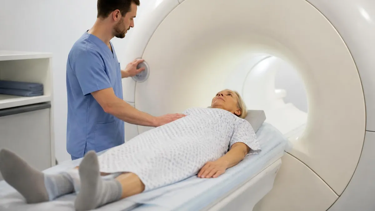
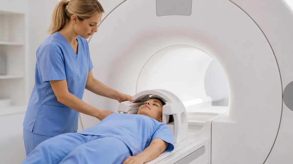
The scan itself is straightforward. You lie on a padded table, usually on your back. A coil (essentially a specialized antenna) is placed over your pelvis and strapped down loosely. You will be given earplugs or headphones because the scanner is loud, somewhere between a jackhammer and a dot matrix printer from 1992.
The table slides into the magnet bore, and the technologist talks to you through an intercom from the control room. You will hear sequences of buzzing and tapping that change as the machine runs different pulse sequences. Each sequence lasts a few minutes. You have to stay still; even small movements blur the images and may force the technologist to repeat a series.
Absolute and relative contraindications include: most cardiac pacemakers and implanted defibrillators (newer MR-conditional devices may be allowed under strict protocol), cochlear implants, certain aneurysm clips, implanted neurostimulators, metallic foreign bodies in the eye, and the first trimester of pregnancy (caution rather than absolute). Always tell the technologist about any implant, however small, before entering the magnet room. If you are claustrophobic, ask whether the facility offers a wide-bore or open MRI scanner and whether oral sedation can be arranged in advance.
Most pelvic MRI exams take 30 to 45 minutes. Multiparametric prostate MRI can run 45 to 60 minutes because it includes diffusion-weighted imaging (DWI) and dynamic contrast-enhanced (DCE) sequences.
Rectal cancer staging protocols often take 30 to 40 minutes plus high-resolution oblique sequences perpendicular to the tumor axis. MR enterography for IBD can add another 30 minutes for small bowel imaging. None of this hurts.
The biggest complaints we hear are noise, the temptation to sneeze at the wrong moment, and the fact that you cannot scratch an itch for 40 minutes. Some patients fall asleep.
When the scan ends, the table slides out, the coils come off, and you are free to go. There is no recovery period for routine MRI. If you received an antispasmodic injection, your vision may be slightly blurred for an hour or so, so arrange a ride if you are sensitive to those medications.
If you received contrast, drink extra water for the rest of the day to help your kidneys clear the gadolinium. Side effects from gadolinium are rare but include nausea, headache, and very rarely a hypersensitivity reaction. Long-term gadolinium retention is an area of ongoing research; current guidance from regulators is that the benefit of a properly indicated contrast MRI continues to outweigh the theoretical risk in patients with normal renal function.
Pre-MRI Checklist for Patients
- ✓Read and complete the MRI safety screening form thoroughly
- ✓Tell the technologist about every implanted device, even old ones
- ✓Remove all jewelry, watches, hearing aids, dentures with metal
- ✓Mention pregnancy or possible pregnancy before entering the magnet room
- ✓Follow fasting and bowel prep instructions exactly
- ✓Bring a list of current medications and any prior imaging on CD
- ✓Plan for 60-90 minutes door to door, longer if contrast is given
- ✓Arrange a ride if you receive sedation or an antispasmodic injection
Now let us talk about the cost, because this is the question most patients actually want answered and the one that no one in the imaging suite has time to discuss properly.
In the United States, a pelvic MRI without contrast typically lists between $1,000 and $3,500 at hospital-based centers. With contrast, the price jumps to anywhere from $1,500 to $5,000. Free-standing imaging centers are usually cheaper than hospital outpatient departments, sometimes dramatically so.
Cash-pay rates at independent imaging centers can run $400 to $800 in some metro areas, while the same study at the academic medical center next door bills out at $4,000. That kind of spread is hard to believe until you call around yourself.
Most insurance plans cover medically necessary MRI but require prior authorization. That paperwork takes anywhere from a day to two weeks. Skip it and the claim will be denied even if the test was medically appropriate.
High-deductible plan members often owe the full negotiated rate until the deductible is met, which is why so many people compare cash prices across imaging centers. The federal price transparency rule means hospitals must publish negotiated rates online, though they are notoriously hard to find.
If cost is a concern, call several imaging centers and ask for the cash price for CPT code 72195 (MRI pelvis without contrast), 72196 (with contrast), or 72197 (without and with contrast).
Pelvic MRI Strengths and Limitations
- +Outstanding soft tissue contrast, especially for prostate, uterus, ovaries, rectum
- +No ionizing radiation, safe to repeat over time
- +Multiparametric capability (T1, T2, DWI, DCE) allows tissue characterization
- +Detailed roadmap for surgical and radiation oncology planning
- +MR arthrography sees labral tears that X-ray and CT miss
- +Reproducible across centers when standard protocols are followed
- −Expensive and often requires prior authorization
- −Long scan times, hard for claustrophobic or restless patients
- −Contraindicated with many implanted devices
- −Motion artifact from breathing, bowel peristalsis, restless patients
- −Gadolinium contrast carries small risks and is restricted in poor renal function
- −Limited availability and long wait times in some regions
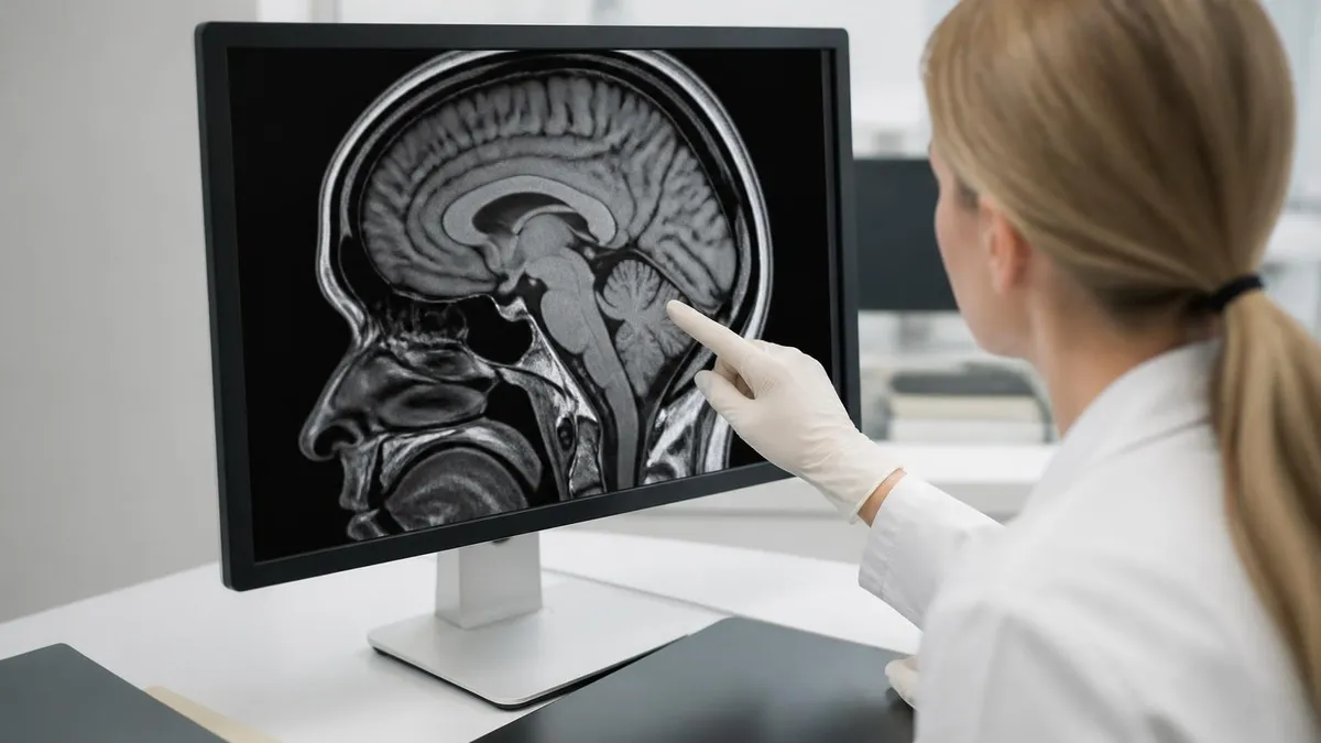
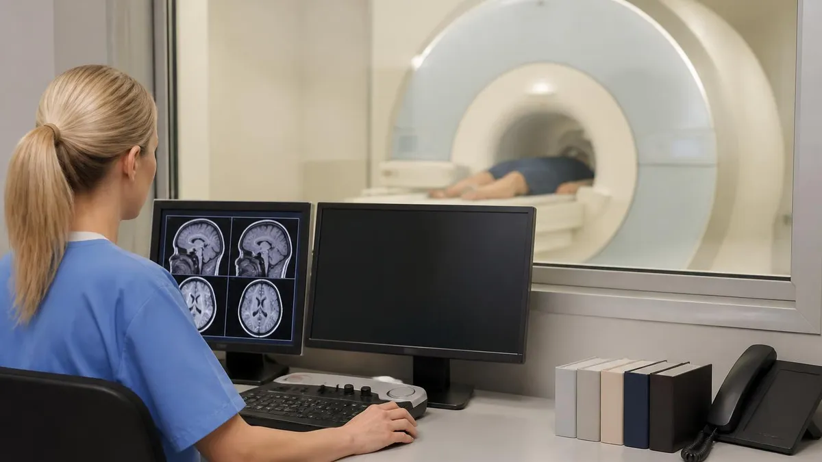
Once the scan is done, the images travel from the scanner to a workstation where a radiologist (typically a body imager, musculoskeletal radiologist, or genitourinary subspecialist depending on the indication) reads them.
Turnaround time depends on the facility and on how urgent the study is. Outpatient scans usually have a report ready within one to three business days. Inpatient and ER scans are often read within an hour.
The report will follow a structured format: clinical history, comparison studies, technique, findings, and impression. The impression is the part your ordering physician will read first. The findings section is the long, organ-by-organ inventory that supports the impression.
Reading your own report can be intimidating because it is written in radiologist language. A few translation tips help.
The phrase "no acute abnormality" means nothing emergent was seen. "Unchanged from prior" is reassuring and means whatever finding exists has not progressed since the last comparable study. "Indeterminate lesion, recommend follow-up" is not a diagnosis; it means the radiologist needs more information, usually another scan in three to six months.
"PI-RADS 3" on a prostate MRI means an equivocal lesion with intermediate probability of clinically significant cancer; PI-RADS 4 means probably significant cancer; PI-RADS 5 means highly likely significant cancer. "BI-RADS" is a similar scoring system but for breast imaging, not pelvic. For rectal cancer, you will see T1 through T4 staging based on depth of invasion and N staging based on involved lymph nodes.
It is normal to leave the radiology section with more questions than you started with. The report is a snapshot, not a treatment plan. Always bring it to your referring physician, who will combine the imaging with your symptoms, labs, and exam to decide what comes next.
If you are a patient, do not Google the unfamiliar terms until after that conversation. If you are a tech student, the report is where your protocol choices show up in plain English: every weighted sequence you ran, every plane you angled, every contrast bolus you timed, all of it is feeding into those findings.
MRI Questions and Answers
If you are sitting an MRI registry exam, pelvic imaging shows up in a surprising number of questions, partly because the anatomy is dense and partly because the protocols are so indication-specific.
Know the standard sequences: T1-weighted axial for fat and post-contrast comparisons, T2-weighted in three planes for high-resolution anatomy, fat-suppressed T2 or STIR for edema and inflammation, diffusion-weighted imaging for cellularity, and dynamic contrast-enhanced sequences for perfusion.
For prostate, know that PI-RADS uses T2, DWI (including high b-value images and ADC maps), and DCE; that the peripheral zone is assessed primarily on DWI and the transition zone primarily on T2.
For rectal cancer, know the mesorectal fascia, the importance of high-resolution thin-section T2 oblique to the tumor axis, and why DWI helps with restaging after chemoradiation.
For gynecologic MRI, know the layered appearance of the uterus on T2 (junctional zone, myometrium, endometrium), the appearance of mature teratomas (fat saturation drops the signal), and the kissing ovaries sign of deep endometriosis.
Beyond the registry, remember the patient on the other side of the magnet. Forty-five minutes is a long time to lie still while a giant machine bangs around your head.
A few extra minutes spent explaining the noise, the contrast injection, the breath-hold sequences, and what happens after the scan turns a stressful test into a tolerable one. Patients who understand what they are getting move less, finish faster, and produce diagnostic images.
That is true on the registry exam and it is true in real life. The best radiology departments treat patient communication as part of the protocol, not as something to do after the imaging is done. The technologist who takes thirty seconds to explain why the room is so loud will save five minutes of retakes later.
Pelvic MRI Final Pre-Scan Checklist
- ✓Ask if the imaging center will share images on CD or via patient portal
- ✓Memorize your panic button position before the table moves in
- ✓Tell the tech if any prior MRI caused anxiety or claustrophobia
- ✓Ask which body coil will be used and how it will be positioned
- ✓Confirm whether the radiologist reading is a body or MSK subspecialist
- ✓Schedule a follow-up appointment with your referring physician within 5 days
One last reminder before you walk into your first pelvic MRI. Ask the imaging center if they will share the images on a CD or via a patient portal. Having your own copy makes second opinions and future comparisons far easier.
And do not be embarrassed to ask the technologist to slow down, to give you the panic button you can squeeze, or to explain what is happening at each stage. Good imaging departments expect those questions and welcome them. The patient who understands the process moves less, finishes faster, and ends up with cleaner images that need no retakes.
About the Author
Medical Laboratory Scientist & Clinical Certification Expert
Johns Hopkins UniversityDr. Sandra Kim holds a PhD in Clinical Laboratory Science from Johns Hopkins University and is certified as a Medical Technologist (MT) and Medical Laboratory Scientist (MLS) through ASCP. With 16 years of clinical laboratory experience spanning hematology, microbiology, and molecular diagnostics, she prepares candidates for ASCP board exams, MLT, MLS, and specialist certification tests.