Computed Tomography Test 2026 June
Boost your Computed Tomography exam score with practice questions and detailed answer explanations. Track progress with instant feedback.
Computed Tomography Test Questions and Answers
A process that creates a succession of in-depth images of various body parts using an X-ray machine connected to a computer. Various angles of photographs are used to see tissues and organs in three dimensions.
The invention of CT dates to 1972.
A computed tomography scan is a type of medical imaging procedure used to produce in-depth pictures of the body’s inside.
The purpose of a CT scan of the thorax is to map out the size, shape, and location of the organs in the upper abdomen and chest. To inspect the lungs or other places for bleeding or fluid accumulations, check for chest inflammation or infection, and search for lung blood clots.
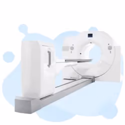
In order to create cross-sectional (slices) images inside certain body regions from different angles, CT scans—medical imaging procedures—use ionizing radiation.
A narrow X-ray beam is quickly spun around a patient’s body during a procedure known as “computed tomography,” or CT. This produces signals that are then analyzed by the machine’s computer to create cross-sectional images, or “slices,” of the patient’s body.
As of May 1st, 2026, the average compensation for a technologist performing computed tomography (CT) scans in the United States is $78,800, but the range frequently varies from $71,980 to $86,140.
While there may be a little increase in your risk of cancer at far greater levels, the modest radiation doses used in CT scans have not been proven harmful over the long term. The numerous advantages of CT scan far outweigh any minimal possible risks.
The skills required of a CT scan technologist include reading a doctor’s scanning orders correctly, giving contrast agents, setting up and using the CT scanner, and positioning the patient to get the best results.
A computerized axial tomography scan can be utilized to aid in the diagnosis of illness, the planning of treatment, or to assess the efficacy of that treatment.
Digital tomography Angiograms, or the imaging of arteries and veins throughout the human body, are performed using angiography computed tomography technology.
In many different parts of the body, CT scans can be used to detect disease or damage.
A medical imaging method called cone beam computed tomography uses divergent X-ray computed tomography to create a cone-shaped image. In various fields, including implant dentistry, ENT, orthopedics, and interventional radiology, CBCT has grown significantly for treatment planning and diagnosis.
A process that creates a succession of finely detailed images of locations inside the body using a computer connected to X-ray equipment that emits a very low dosage of radiation. Several angles of photography were used to construct 3-D views of tissues and organs. According to their age and smoking history, adults with a high risk of developing lung cancer are advised to undergo low-dose computed tomography as a screening test. Also known as a low-dose CT scan and LDCT.
Gamma rays are used in the nuclear medicine tomographic imaging technology known as single-photon emission computed tomography. While using a gamma camera is extremely comparable to traditional nuclear medicine planar imaging and can produce full 3D data.
It was invented by EMI Laboratories’ British engineer Godfrey Hounsfield and Tufts University’s South African-born physicist Allan Cormack.
- Determine whether you want to pursue a profession as a CT scan technician. As a CT scan technologist, your responsibilities often involve setting up and running diagnostic equipment, maintaining patient records, and arranging patients for clear CT scan images. The process must be repeated if the image needs to be clarified or clarified. You thoroughly discuss the process and address any patients’ queries before they undergo a CT scan. You are qualified to do ultrasounds and other radiology treatments since you are a certified radiologic technologist.
- High school graduation is a prerequisite for a radiography degree program. A solid math, science, and computer science foundation will help you. Biology and psychology electives could be useful as well.
- You have higher employment prospects if you have an associate’s degree in nuclear medicine technology, diagnostic medical sonography, or radiography. Through these classes, you can learn medical language, healthcare systems, radiology techniques, radiology gear, patient care, and anatomy. A clinical practicum is also a component of such curricula. Contact the community or technical college in your area for information on available programs.
- The American Registry of Radiologic Technologists offers voluntary certification (ARRT, www.arrt.org). Before seeking CT scan certification, you must first achieve ARRT certification in a supporting field, typically radiography, radiation therapy, or nuclear medicine technology. After receiving this initial certification from the ARRT, candidates must complete 16 credits of training in particular CT specialties and verify the supervised clinical practice. These courses can be completed in six months to a year. You can take the ARRT certification exam if you’ve met the prerequisites for clinical training.
- The U.S. Bureau of Labor Statistics reports that each state has its licensing standards. Through the American Society of Radiologic Technologists, you can learn more about the license requirements in your state.

- ✓Review the official Computed Tomography Test exam content outline
- ✓Take a diagnostic practice test to identify weak areas
- ✓Create a study schedule (4-8 weeks recommended)
- ✓Focus on your weakest domains first
- ✓Complete at least 3 full-length practice exams
- ✓Review all incorrect answers with detailed explanations
- ✓Take a final practice test 1 week before exam day
Computed Tomography Practice Test Questions
Prepare for the Computed Tomography Test exam with our free practice test modules. Each quiz covers key topics to help you pass on your first try.
Computed Tomography Test CT Anatomy and Cl...
Computed Tomography Exam Questions covering Test CT Anatomy and Clinical Applications. Master Computed Tomography Test concepts for certification prep.
Computed Tomography Test CT Contrast Media...
Free Computed Tomography Practice Test featuring Test CT Contrast Media and Patient Preparation. Improve your Computed Tomography Exam score with mock test prep.
Computed Tomography Test CT Image Quality ...
Computed Tomography Mock Exam on Test CT Image Quality and Reconstruction. Computed Tomography Study Guide questions to pass on your first try.
Computed Tomography Test CT Radiation Safe...
Computed Tomography Test Prep for Test CT Radiation Safety and Dosimetry. Practice Computed Tomography Quiz questions and boost your score.
Computed Tomography (CT Brain)
Computed Tomography Questions and Answers on (CT Brain). Free Computed Tomography practice for exam readiness.
Computed Tomography (CT)
Computed Tomography Mock Test covering (CT). Online Computed Tomography Test practice with instant feedback.
Computed Tomography Knowledge Questions an...
Free Computed Tomography Quiz on Knowledge Questions and An. Computed Tomography Exam prep questions with detailed explanations.
Computed Tomography Science
Computed Tomography Practice Questions for Science. Build confidence for your Computed Tomography certification exam.
- +Industry-recognized credential boosts your resume
- +Higher earning potential (10-20% salary increase on average)
- +Demonstrates commitment to professional development
- +Opens doors to advanced career opportunities
- −Exam preparation requires significant time investment (4-8 weeks)
- −Certification fees can be $100-$400+
- −May require continuing education to maintain
- −Some employers may not require certification
Computed Tomography Test Questions and Answers
About the Author
Certified Professional Development Expert & Niche Certification Advisor
University of Pennsylvania Graduate School of EducationDr. Alexandra Kim holds a PhD in Professional Studies from the University of Pennsylvania and is a Certified Professional in Learning and Performance (CPLP) and Certified Professional in Talent Development (CPTD). With 17 years of corporate training and professional certification advisory experience, she helps professionals navigate specialized, emerging, and cross-industry certification programs.