FREE Computed Tomography (CT Brain) Questions and Answers
A patient presents with sudden severe headache, often described as "the worst headache of their life." Which type of brain hemorrhage is most likely?
A sudden severe headache, often described as "the worst headache of their life," is a common presentation of subarachnoid hemorrhage, often caused by the rupture of an intracranial aneurysm.
What is the typical Hounsfield Unit (HU) range for normal brain tissue on a non-contrast CT scan?
Normal brain tissue typically has Hounsfield Unit values ranging from -100 to 100 HU on a non-contrast CT scan.
Brain non-contrast CT: Hypertension history and rapid start of left facial droop. What does the picture depict?
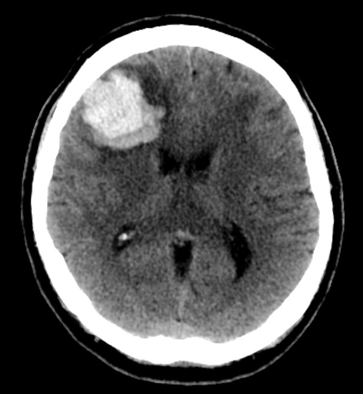
A significant high-density area caused by severe bleeding can be seen in the right frontal lobe in the picture. Since there was no contrast on the CT, this cannot be an enhancing mass.
Its location suggests that it is intra-axial rather than extra-axial in the brain, ruling out subarachnoid and subdural bleeding..
The calcified choroid plexus, a common occurrence, is the focus of high density in the posterior horn of the right lateral ventricle.
What is the primary advantage of using contrast enhancement in brain CT imaging?
Contrast enhancement in brain CT imaging improves the visualization of blood vessels and enhances the differentiation between normal and abnormal tissues.
Colon cancer patient after brain contrast MRI Which diagnosis is more likely?
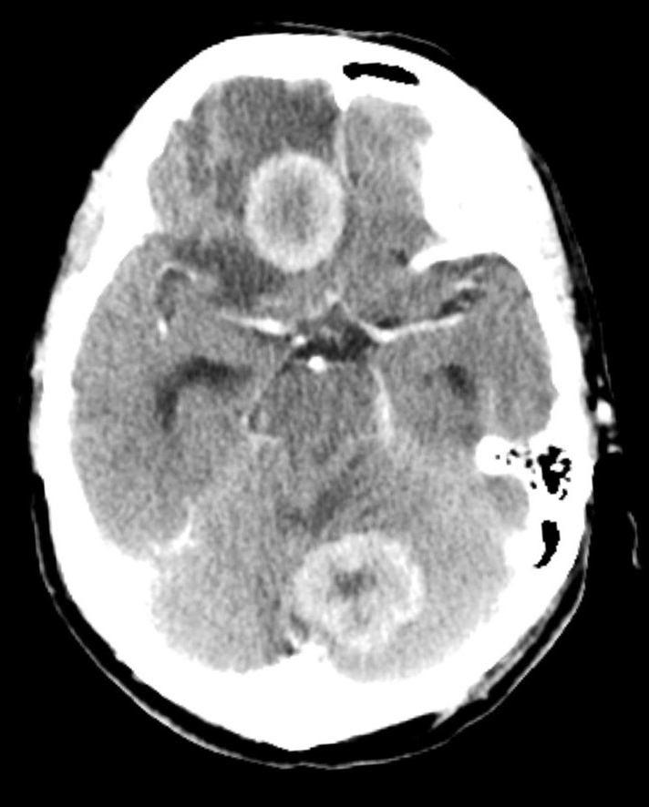
A brain tumour in a patient with a known cancer is almost certainly the result of a metastatic illness. It is uncommon for benign or malignant primary lesions to appear simultaneously.
Both the enhancing masses and normal vessels are shown in the photograph.
There is no evidence of subarachnoid haemorrhage.
A digital radiograph was produced while the patient bed was translated (moved) through the gantry, maintaining the X-ray tube and detector array in place.
A scout image in CT (computed tomography) is a low-dose radiographic image that is acquired before the actual CT scan. It is also known as a localizer or program. The scout image is used to define the exact location of the area of the body that needs to be scanned and to plan the CT scan accordingly.
What is the typical Hounsfield Unit (HU) range for normal brain tissue on a non-contrast CT scan?
Normal brain tissue typically has Hounsfield Unit values ranging from -100 to 100 HU on a non-contrast CT scan.
Which CT scan view provides the best visualization of the interhemispheric fissure?
The coronal view in CT provides the best visualization of the interhemispheric fissure, which separates the two cerebral hemispheres.
Non-contrast CT brain: anticoagulated patient with right hemiparesis and abrupt onset of disorientation. What does the picture depict?
High-density material fills the left lateral ventricle. This is blood which extended from an intracerebral haemorrhage of the adjacent peri-ventricular white matter.
BEAM COLLIMATION and DETECTOR COLLIMATION settings determine the section width in the MSCT System.
MSCT beam collimation is the X-ray beam width used to acquire images during a CT scan. The pre-patient collimation and the post-patient collimation determine it.
Which type of brain imaging is most useful for assessing the presence of calcifications?
CT is excellent for detecting calcifications due to its ability to differentiate dense structures from surrounding tissues.
Which type of intracranial hemorrhage appears hyperdense on a CT scan?
Epidural hemorrhages appear hyperdense (bright) on CT scans due to their location between the inner skull and the dura mater.
Poor historian, his perplexity has grown over the past few weeks. What does the picture depict?
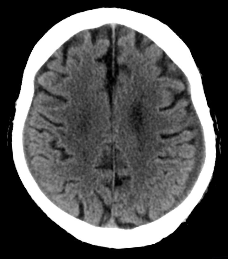
It is easy to see the left-sided subdural collection. On the right, there is a small subdural collection as well
Remember to keep looking at the scan methodically, even if you notice one irregularity.
The quantity of detector rows X Size of each detector is Section Width; for example, 64 x 0.625 is 40mm.
Beam Collimation/Number of Detector Rows = Detector Collimation, where 0.625 = 40 mm/64.
Which type of brain lesion commonly presents as a ring-enhancing lesion on contrast-enhanced CT?
Brain abscesses often present as ring-enhancing lesions on contrast-enhanced CT scans due to the contrast material accumulating around the outer edge of the lesion.
Which brain imaging technique is most sensitive for detecting acute ischemic strokes?
MRI is more sensitive than CT in detecting acute ischemic strokes due to its ability to visualize tissue changes associated with early stroke development.