Radiography

Radiography is a broad field that uses X-rays, MRIs and CT scans to diagnose health conditions. Radiographers are healthcare professionals who facilitate these scanning procedures and ensure that they are performed safely.
The radiographer’s duties include monitoring patients and getting them ready for the scan, assisting the radiologist with the procedure and documenting important information. This job requires a high level of compassion and understanding for patients.
Radiography Salary
Radiography techs are essential members of the healthcare team who use X-rays, CT scans, and mammograms to diagnose patients. They also use advanced technologies such as gamma rays and ionizing radiation to treat diseases.
The salary of a radiography technician can vary depending on a number of factors, including education, experience, certifications, and workplace setting. Radiographers who have more experience and higher credentials tend to make a higher salary than those with less education.
A radiography technician’s salary can increase with additional relevant education, according to the American Society of Radiologic Technologists (ASRT). RAD techs can earn a bachelor’s degree in radiological science or healthcare administration, which can help them advance in their career and earn more money.
Cleveland University-Kansas City’s Associate of Applied Science in Radiologic Technology program provides focused, eight-week evening classes. Students learn hands-on skills through labs, equipment rooms, and clinical experiences at local imaging departments. CUKC’s courses don’t require transfer of general education credits from elsewhere, so students can focus on professional training right away. The University is also accredited by the Joint Review Committee on Education in Radiologic Technology, so graduates will have a solid foundation for their future careers.
Radiography Vs Radiology
Radiography is the process of taking images of different parts of a patient’s body using radiation. It is an important part of medical science and helps doctors in diagnosing diseases effectively.
Radiographers work in the field of medical imaging and they take X-rays, computed tomography (CT scan), and magnetic resonance imaging (MRI) scans. They also use ultrasound to produce clear images of the body’s internal organs and tissues.
Another important role of a radiographer is helping patients with their comfort during scans. These tests involve confined areas and often require long periods of time, so it is important for radiographers to watch and monitor patients carefully to ensure their safety and comfort.
The terms “radiography” and “radiology” are commonly confused, but they have distinct meanings and require a number of different educational qualifications and training requirements. A radiographer completes an associate degree, while a radiologist earns a bachelor’s or master’s degree in medical imaging or a related field.
Dental Radiography
Radiography is the process of taking pictures of your teeth and jaws with X-rays. These X-rays allow dentists to see what’s going on inside your mouth and to detect any problems that may not be visible to the naked eye during a visual exam alone.
Dentists typically use a variety of dental X-rays to assess the health of your teeth and gums. These X-rays may include bitewing, periapical (also called ‘around the apex’) and panoramic radiographs.
Bitewings are taken to help your dentist spot signs of decay between your back teeth, or bicuspids (teeth in front of the molars). Periapical X-rays show your entire tooth from the crown to the root tip, so your dentist can see any changes that occur beneath the gum line.
Panoramic X-rays are often used to diagnose issues such as cysts or jaw disorders. They also help evaluate the status of your teeth and jawbone, which can be helpful for orthodontic treatments like braces.
Digital Radiography
Digital radiography, or DR, is an advanced imaging technology that produces clear, high-quality images without the use of film. Compared to conventional X-rays, this method is much safer and provides greater image quality.
Digital X-rays are also faster and easier to produce, which can save patients time. They’re also environmentally friendly, because they don’t require chemicals to develop them. They can also be transferred to other facilities or patients who request an individual copy.
This type of technology is also more affordable than other alternatives, which means health care professionals can afford to offer it to their patients. It’s also faster, making it more convenient for patients to schedule appointments and diagnostic tests.
In addition to a wide range of medical applications, digital radiography is used for aerospace product examination and casting and welding inspection. It can help companies ensure that their products are safe and reliable, and it can also improve productivity by eliminating the need to transport physical X-ray films between departments.
Radiography Definition
Radiography is the medical science of using high-energy radiation (X-rays) to produce images of tissues, organs and bones inside the body. These radiographic images are used to diagnose medical conditions and treat patients with various treatments.
Radiographers are health care professionals who operate X-ray equipment to create diagnostic images for use by radiologists, physicians with advanced training in reading these images. They may eventually specialize in certain areas of imaging such as MRI, CT scans and sonography.
Typically, the radiographer receives orders from the radiologist for the type of scan that a patient needs and then operates the equipment necessary to produce the images. It is important that the radiographer ensures that only the amount of radiation needed to produce a quality diagnostic image is applied.
A radiographer may work in a hospital or other health care facility, or in a private practice setting. They must complete a program in radiography that is accredited by the Joint Review Committee on Education in Radiologic Technology in order to earn certification from the American Registry of Radiologic Technologists (ARRT).

Radiography Degree
Radiographers work in hospitals and medical facilities, providing imaging services to help doctors diagnose and treat patients. They use a variety of forms of radiation, including X-rays and magnetic resonance imaging (MRI), to take images of the body’s structures.
Radiography is one of the fastest-growing healthcare fields. It is growing in response to the need for noninvasive imaging technologies and procedures.
As a radiographer, you can expect to enjoy a rewarding career that helps people get better. You will often form personal relationships with your patients, and you can become involved in their treatment.
The field is growing faster than many others, and jobs for radiographers are expected to increase by 6.3% through 2031.
A degree in radiography can help you get started in a career in this field. An associate degree may be sufficient for entry-level positions, but earning a bachelor’s can enhance your abilities and give you the training you need to advance to management or supervisory roles.
You should look for a college or university that offers an accredited program in radiologic technology. Accreditation is a validation process that makes sure schools offer a quality education. It also impacts your eligibility for licensure, credentialing exams and the ability to transfer credits from one program to another.
Radiography Technician
Radiography technicians use medical imaging equipment to produce X-rays and other diagnostic images of the human body. Their precise work allows doctors to diagnose and treat a wide variety of diseases.
Typical duties of a radiology technician include preparing patients for radiologic examinations, positioning patients, and capturing clear, usable images. They also monitor patients’ safety and calibrate the equipment.
Radiographers may be called on to inject patients with barium- and iodine-based contrast agents to enhance images. They also perform procedures to guide catheters, vena cava filters, and stents through veins or arteries.
Education requirements for radiography technologists typically include an associate degree or certificate in radiologic technology. Most programs are two years in length and provide both clinical and classroom instruction.
Radiologic technologists generally work in hospitals, diagnostic laboratories, physicians’ offices, and other health care facilities. Salaries vary depending on location and employer type.
Medical Radiography
Radiography is the process of using x-rays and other forms of radiation to create an image of the body. These images are used by medical professionals to diagnose and treat illnesses or injuries.
Radiographers are trained to use a variety of imaging devices such as x-rays, computed tomography (CT) scans, magnetic resonance imaging (MRI), ultrasound and nuclear medicine to examine patients. They also provide patient education, help patients prepare for tests and ensure that the equipment produces accurate results.
X-rays are a form of radiography and can be used to detect bone fractures, certain tumors and abnormal masses, pneumonia, some types of injuries, calcifications, foreign objects or dental problems. They can also be used to monitor the growth of breast cancer and to detect tiny bits of calcium called microcalcifications that appear as bright specks on mammograms.
Medical radiography is an excellent career choice for people interested in a scientific discipline and helping to improve patient care. It is a highly competitive field with a high median salary and a wide range of career options for radiographers.
Radiography Exam Questions and Answer
An imaging technique called radiography uses X-rays, gamma rays, other ionizing radiation, and non-ionizing radiation to see an object’s internal structure. Medical and industrial radiography are two examples of radiography’s applications. Airport security employs similar procedures.
The median pay for them, according to the U.S. Bureau of Labor Statistics, is $65,140.
When determining which medical imaging technique is more challenging, sonography or radiography, it is imperative to consider the intricacy of both types of imaging procedures. Sonography involves visualizing complex anatomy with high-frequency sound waves, while radiography requires producing an image by exposing a patient to radiation. Both require extensive training and practice for successful outcomes and appropriate diagnoses. Sonography can be more difficult than radiography in some circumstances due to the level of detail required. While radiology can provide excellent sharpness in images of bones or soft tissues, sonographers must pay special attention to even miniscule details while imaging organs such as fetuses or blood vessels in order to provide accurate readings.
The processes used in fluoroscopy and radiography appear to be similar. Fluoroscopy, on the other hand, produces moving images of the interior of the body, whereas radiography produces a static image of the internal structure of a body using gamma rays.
There is a difference between radiography and radiology. With the aid of sophisticated tools like X-ray machines and MRI scanners, radiography creates inside images of the human body. The branch of medicine known as radiology employs radiologic imaging methods to identify illnesses and disorders.
- radiographer.
- x-ray operator.
- sonographer.
- clinical photographer.
- medical instrument technician.
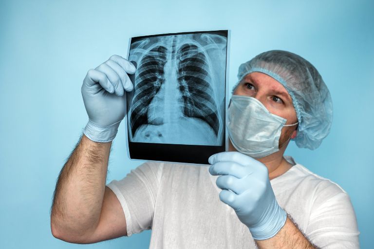
The purpose of a chest radiograph, often known as a chest X-ray or chest film, is to diagnose illnesses that affect the chest, its contents, and adjacent structures.
The capacity of an imaging system to tell two nearby structures apart from one another is known as spatial resolution.
Digital radiography (DR) is a more sophisticated method of X-ray inspection that quickly generates a digital radiographic image on a computer.
The pay is reasonable. Your earning potential will be influenced by how quickly you advance through a hospital’s grades and how much private work you take on. It can be quite profitable if you work a lot “on call” or specialize in something like sonography or ultrasound.
Radiography is an X-ray procedure that bears the same risks as other X-ray procedures.
Computed radiography (CR), a computerized alternative to traditional X-ray film radiography, has many advantages for inspection duties because it essentially eliminates consumable use and substantially reduces image production time.
A radiography degree can be very demanding. Although there is a lot of material to get through, it is not very difficult to learn.
If you complete additional courses or training, a radiology degree can be used to become an ultrasound technician. A medical sonography license may also be required by some states or employers. Radiologists must acquire the skills necessary to handle diagnostic sonography equipment since the imaging technology is different.
Depending on what the state or employer requires. Having a bachelor’s degree in radiation therapy or a similar subject may be preferred by some companies, while an associate’s degree may be acceptable by others. Radiation therapists must, however, be licensed or certified, which frequently entails completing a national certification exam. As a result, if you already have a radiography degree, you might need to complete extra coursework or training.
Yes, you may earn a Ph.D. in radiography. In radiography or related subjects like radiology or medical imaging, certain universities offer Ph.D. programs. Research projects on subjects like MRI, PET, artificial intelligence, or patient-centered care may be part of these programs, which typically call for an undergraduate degree in radiography or a closely related profession.
Patient radiation exposure from digital X-rays is 90% lower than that from traditional X-ray machines.
Despite the fact that ZipRecruiter reports monthly incomes as high as $10,458 and as low as $2,208, the majority of radiography salaries in the US are currently between $4,583 (25th percentile) and $8,416 (75th percentile).
The study of radiography necessitates a high level of mathematical expertise.
In computed radiography, a particular phosphor coating stores the radiation’s energy when imaging plates are subjected to X-rays or gamma rays. The latent picture is then extracted from the plate using a scanner, a specialized device that stimulates the plate with a highly precisely focused laser beam.
With this method, data is recorded while an object is being examined using X-ray-sensitive plates, and it is then instantly transferred to a computer without the necessity of an intermediate cassette.
Digital X-rays, as opposed to traditional X-rays, offer better, clearer images with high resolution that may be enhanced or altered as necessary, reducing the need for repeating the procedure. Traditional X-rays cannot be manipulated since they are printed on a film.
A photostimulable phosphor plate, or PSP for short, is used in indirect systems to identify X-rays and record the image. The image is then sent to the computer using a USB cable or a network connection after being scanned in a PSP scanner.
It works by covering the esophagus, stomach, or intestine with a substance that is not absorbed by the body, allowing X-rays or CT scans to clearly show diseased or damaged areas.
During an exposure, phosphor crystals take in and momentarily retain the x-ray energy, creating a latent image.
It can normally take up to 13 years to become a radiologist.
Degree programs last three to four years when taken full-time or up to six years when taken on the side.
The average annual salary for a Medical Radiography in the United States is $66,133 as of May 13, 2023.
In the USA, radiographers typically make $65,813 a year.
There is a ton of physics in radiology, but you won’t need to know much of it to practice on a daily basis, even though you’ll internalize a lot of it. The practical (and less practical) aspects of imaging physics are extensively tested on the radiology board exams.
Depending on the radiation source and equipment being utilized, various formulas are accessible for figuring out the exposure period in radiography. The following is a typical formula for radiographic exposure time: Exposure Time (minutes) = Film Factor x Source to Film Distance^2 / Roentgen per Hour at 1 Meter
HVL is expressed in aluminum millimeters. Because the filtered photons have more energy after being filtered by one HVL, the succeeding HVL will be greater (thicker material is required to attenuate half of the penetrating beams). The following formula relates it to the linear attenuation coefficient (): HVL = 0.693 / μ
The Source to Film Distance, or SFD, is the measurement of the distance between the radiation source and the film along the direction of the beam. SOD and the Object to Film Distance (OFD) can be combined to get the SFD using the following formula: SFD = SOD + OFD
- Cut a rectangular piece of material longer than the film width and wide enough for the number of steps.
- Mark the material into equal segments along the length for the number of steps.
- Cut each segment at an angle to create a constant thickness difference between steps.
- Glue or tape the segments together to form a step wedge with the thinnest segment at one end and the thickest segment at the other end.
- Label each step with its thickness and number.
- Place the step wedge on a film and expose it to radiation to test your exposure parameters. Compare the image with a standard density chart or use a densitometer to measure the optical density of each step.
The head of the patient is supported by the chin, forehead, and side rests. For a clearer view, the patient could be given a bite blocker to gently open the mouth.
There are several ways to reduce patient dose in radiography, depending on the type of radiographic examination and the equipment used. Some of the common methods are:
- Use fast film or digital detectors that require less radiation exposure to produce an image.
- Use rectangular collimation to limit the radiation beam to the area of interest and reduce scatter radiation.
- Use constant potential X-ray units that produce more uniform radiation output and allow lower exposure settings.
- Use longer source-to-film distance to reduce the intensity of radiation reaching the patient and the film.
- Use rare-earth filtration to remove low-energy photons that contribute to patient dose but not image quality.
- Use low-dose protocols and new image-processing techniques that can enhance image quality while reducing radiation exposure.
- Use appropriate exposure parameters (kV, mA, time) for each patient and examination type, and avoid unnecessary or repeated exposures.
- Use shielding devices such as lead aprons, thyroid collars, and gonad shields to protect radiosensitive organs from scatter radiation.
To study radiography, you need to have a pre-university qualification in science subjects, such as physics, chemistry, biology, and mathematics. You can then apply for a radiography and medical imaging course at a university or a diploma programme at a college. The duration of the course may vary from two to four years, depending on the level and type of qualification.
MRI is a type of radiography; however, unlike X-rays, which carry health hazards with repeated exposure, MRI does not employ ionizing radiation.
Being a radiology technician carries risks, such as radiation exposure and a higher risk of developing cancer. However, with time, threats like cancer development may become less dangerous if safety rules are correctly followed, and contemporary technology is used.
Radiography is not boring; however, it may not be for everyone. Utilizing medical imaging to aid in the diagnosis and treatment of various diseases and ailments is a key component of the dynamic and gratifying field of radiography. Radiographers work with multiple patients and imaging technologies, including CT, MRI, X-ray, and ultrasound. Additionally, they work along with other medical specialists like radiologists, doctors, nurses, and physicists.
The Radiography program’s professional phase admission is competitive. If accepted, the candidate can start the professional phase the following summer.
Unlike nursing school, radiology tech school is not more difficult. Contrary to popular belief, nursing school is not more difficult than radiology tech school. In fact, radiology tech school and nursing school are both challenging in their own ways and call for various knowledge and skill sets.
Radiographers are likewise in high demand, not just in the United States but also internationally.
Sonography and radiology employ different technology. Radiology technicians may use radiation-emitting equipment such as X-rays, CT scans, or magnetic resonance imaging (MRIs). High-frequency sound waves are used by diagnostic medical sonographers to produce their images.
If you are interested in medical imaging and want to assist individuals with their healthcare problems, radiography may be worthwhile. In the rewarding field of radiography, you can utilize your technical expertise to create images of the human body for both therapeutic and diagnostic purposes. Additionally, there are numerous career pathways and chances for progression and specialization in radiography.
Ultrasound is a sort of radiography that creates images of the body’s internal structures by using high-frequency sound waves.
In conventional radiography, the contrast is influenced by the size of the grains, the length of time that they are allowed to develop, the temperature and concentration of the developing solution, and the overall film density.
The most common indications for X-rays are the exclusion of fractures following trauma, the assessment of joint or spinal disease, and the assessment of cardiac disease.
To get a high-quality X-ray image, the right doses are chosen using the exposure parameters. Peak kilovoltage (kVp), milliampere (mA), exposure time (s), and the patient’s distance from the X-ray source are some of the exposure factors (SID).
Digital radiography has many advantages, such as:
- Better image quality: Using software tools and algorithms, digital images can be enhanced, manipulated, and optimized.
- Faster processing and diagnosis: Digital images can be instantly displayed, transmitted, and shared via electronic devices or networks with other health professionals.
- Lower radiation dose: Digital detectors require less radiation exposure and have a wider dynamic range than film, reducing the need for retakes.
- Easier storage and access: Digital images can be stored electronically in databases or cloud systems and integrated with electronic health records or other information systems.
- Plain X-rays or Radiography.
- CT scanning, also known as computed tomography.
- Fluoroscopy, which creates images of an organ that moves.
- Mammography, which is a breast X-ray.
- Angiography, which is a blood vessel x-ray.
Digital radiography has a few drawbacks, but the cost of upgrading old radiographic equipment is perhaps the biggest.
In comparison with conventional X-ray images, such as chest X-Ray, CT images of internal organs, bones, soft tissue, and blood arteries offer higher clarity and detail.
Intraoral radiography uses two kinds of digital imaging systems: computed radiography (CR) and direct radiography (DR).
The distance between the subject to be radiographed and the film, as well as the distance between the tube’s focal point and the film, both have an impact on magnified distortion.
The term “shape distortion” describes how the target object is elongated or shortened. The X-ray tube’s technical and/or structural flaws, inappropriate angulation of the image receptor or axis, or both might cause this look.
Variations in photon concentration, thermal activity inside an electronic device, fluorescent lights, or the sensitivity of the image receptor are all factors that might cause image noise.
The International Commission of Radiological Protection has suggested Diagnostic Reference Levels (DRLs) as a tool for optimization. DRLs show whether a radiographic procedure’s dose to patients or the quantity of radiopharmaceuticals used for imaging is exceptionally high or low.
The abbreviation MSP stands for Mid Sagittal Plane Extraction from Brain MRI.
Superimposed in radiography means that two or more structures are overlapping or lying on top of each other in the same plane of the radiographic image.
With systems that have a moving grid assembly (sometimes referred to as a Bucky device) that oscillates throughout the exposure to blur the grid lines, low frequency grids are used.
A controlled area in radiography is an area that has been designated by an employer to assist in controlling and restricting radiation exposures. A controlled area is usually marked by signs, barriers, or other means to indicate that special procedures are required for people entering the area.
A ghost image occurs when an item or anatomic structure is positioned between the X-ray source and the center of rotation and has a density sufficient to attenuate the X-ray beams.
The height of the lead strip to interspace distance is measured by the grid ratio, which is a reliable indicator of the selectivity of primary to scatter transmission.
Automatic Exposure Control is a device that terminates X-ray exposure.
“As low as reasonably achievable” is abbreviated as ALARA. ALARA stands for abstaining from radiation, especially at low doses, that does not directly benefit you.
An axial projection is equivalent to a central beam that traverses the body’s long axis.
The image receptor could be a normal film screen, a charged electron device for digital radiography, or a photosensitive phosphor plate for computed radiography.
Analog radiography is a type of radiography that uses film or chemical processing to capture and store images of the body’s internal structures.
Attenuation is the reduction of an X-ray beam’s intensity when it passes through matter.
A branch of radiography known as cardiac interventional radiography employs minimally invasive, image-guided techniques to treat heart and blood vascular conditions without surgery.
In conventional and computed radiography (CR), cassettes are rigid holders that are utilized for the image plate and screen film system, respectively.
Following the introduction of digital radiography, collimation—the process of modifying the principal field of ionizing radiation—has reportedly been noted to vary among radiographers.
Contrast radiography is a technique for examining organs that combines the use of X-rays with the delivery of a unique dye known as a contrast medium.
The amount of film darkening is gauged by radiographic density, often known as optical, photographic, or film density. Since it is a measurement of the amount of light transmitted through the film, the term “transmitted density” should technically be used when referring to transparent-base films.
Dental radiography is a type of radiology that uses X-rays to capture images of the teeth and gums. Dental radiographs can help dentists to detect problems like cavities, tooth decay, and impacted teeth.
Deviation Index (DI) values in digital radiography give the radiographer feedback on the suitability of certain exposures, but they can also be monitored as part of a departmental quality assurance program.
A branch of radiology called diagnostic radiography uses imaging methods, such X-rays, to identify diseases and injuries inside the body.
X-ray imaging is referred to as digital radiography. It creates enhanced computer photographs of your dental anatomy using digital X-ray sensors.
The use of greater radiographic exposures—and subsequently doses to the patient—for the same X-ray examination and projection in digital planar radiography is known as dose creep in radiography. Digital radiography systems offer a wide dynamic range and may provide good images even with overexposure, which makes dose creep possible.
Dosimetry’s function is to estimate the dose of radiation an individual will get during a radiological examination.
The tonal difference between an image’s lightest light and darkest dark is known as the “dynamic range.”
EI is a measurement of radiation in the relevant image region on the receptor. It depends on the region of interest (ROI) that the digital radiography workstation has selected for the sort of examination, image processing, and exposure that will be employed.
The degree to which a light-sensitive substance can be overexposed or underexposed and still produce an acceptable outcome is referred to as exposure latitude.
The term “exposure time” describes how long it takes to create an X-ray.
Film speed in radiography refers to the sensitivity of the film to the radiation exposure. The higher the film speed, the less radiation exposure is needed to produce an acceptable image.
The focal spot is where the X-rays come from when making a radiograph.
In radiography, the focal spot size refers to the region of the anode surface that receives the electron beam from the cathode and generates X-rays.
The practice, interpretation, and reporting of radiographic tests and procedures that are required for legal proceedings or law enforcement is known as forensic radiology.
General radiography is the specialty within medical imaging that uses X-rays to examine anatomy. It is also known as plain film radiography because it produces two-dimensional images on a film or a digital detector.
Image acquisition in radiography is the process of capturing an X-ray image using a digital detector. Image acquisition involves selecting the appropriate exposure factors, positioning the patient and the detector, and activating the X-ray tube.
Image blur in radiography is the loss of sharpness or detail in an image, resulting in poor spatial resolution.
Image quality in radiography is the degree to which an image accurately represents the anatomic area of interest and provides sufficient information for diagnosis.
To show defects that are invisible to the naked eye, industrial radiography employs X-ray and gamma radiation.
Radiography is a fascinating field that combines science, technology, and art to produce images of the human body and other objects. Radiography can reveal hidden structures and abnormalities that are not visible to the naked eye. Radiography can also help diagnose and treat various diseases and injuries using minimally invasive procedures.
A tool for displaying a radiograph’s quality is called an “IQI” or “Image Quality Indicator.”
For the accreditation of conventional and online educational programs in radiography, radiation therapy, magnetic resonance, and medical dosimetry, the United States Department of Education (USDE) has solely recognized the JRCERT.